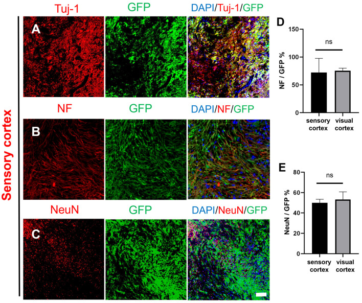
Figure 2.
Histological images of transplanted GFP-labeled NSCs in the injured macaque sensory cortex. (A–C) Transplanted NSCs (GFP, green) differentiated into mature neurons in the injured area of the macaque sensory cortex (Tuj-1/NF/NeuN, red); scale bar, 100 µm. (D) NeuN expression was not different between the sensory cortex and visual cortex, and (E) NF expression was not different between the sensory cortex and visual cortex; ns denotes p > 0.05, means there was no significant difference.

