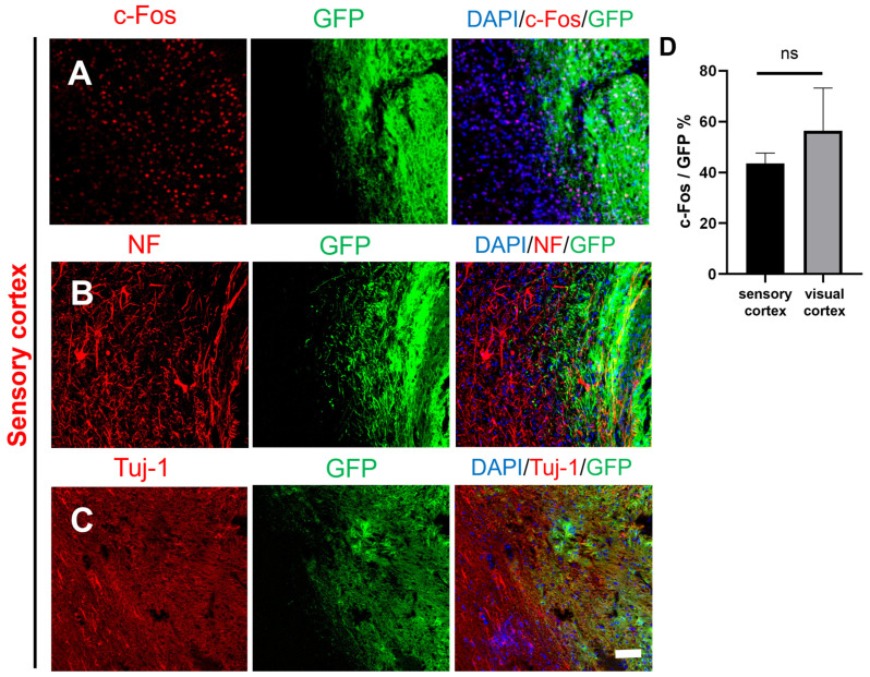
Figure 5.
Histological images of transplanted GFP-labeled NSCs in the border between the graft and host tissue in the sensory cortex. (A) The transplanted NSCs (GFP) differentiated into active cells (DAPI, blue; GFP, green; c-Fos, red). (B–C) The transplanted NSCs (GFP) differentiated into mature neurons at the graft edge (DAPI, blue; GFP, green; Tuj-1, red; NF, red); scale bar, 100 µm. (D) c-Fos expression was not different between the sensory cortex and visual cortex; ns denotes p > 0.05.

