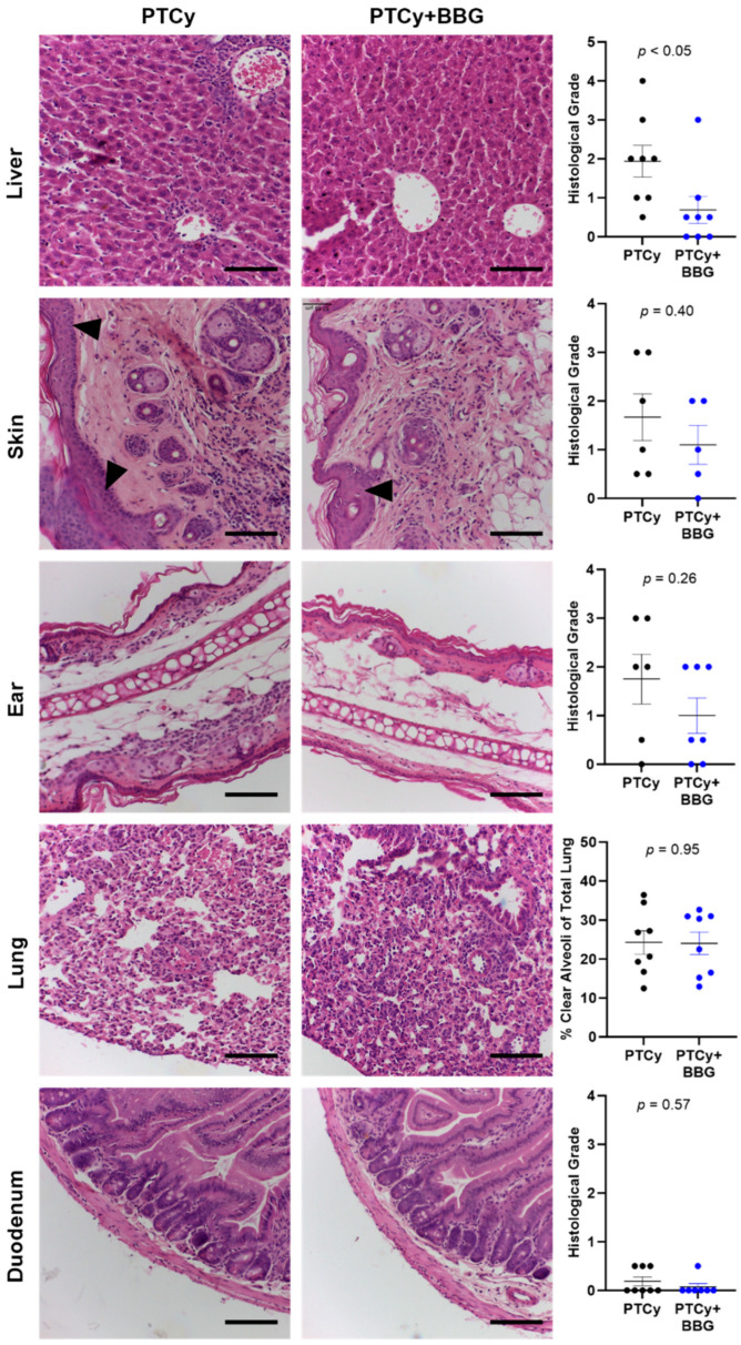
Figure 2.
PTCy with BBG reduces liver GVHD compared to PTCy alone at endpoint. Haematoxylin and eosin-stained liver, skin, ear, lung, and duodenum tissue sections from treated humanised mice at endpoint (Figure 1) were examined for evidence of histological GVHD. Histological GVHD in the liver, skin, ear, and duodenum was assessed using a standardised grading system. Histological lung GVHD was determined as the percent of clear alveoli area of total lung area. Arrowheads indicate epidermal thickening in the skin. Images representative of 5–8 mice per treatment group. Scale bars represent 100 µm. Data are presented as mean ± SEM. Symbols represent individual mice.

