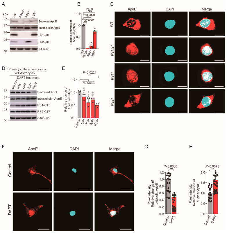
Figure 2.
Presenilin Is Essential for ApoE Secretion, a Novel Role of Presenilin Involved in Alzheimer’s Disease Pathogenesis (Section 4.2). (A) Immunoblot analysis of ApoE secretion and intracellular ApoE, presenilin (PS) 1-CTF, and PS2-CTF expression in WT, PS1/2−/−, PS1−/−, and PS2−/− fibroblasts cultured for 48 h in serum−free medium. (B) Quantification of ApoE secretion from the experiment shown in (A). n = 3. ** p < 0.01, *** p < 0.001. NS, Not significant, by one−way ANOVA followed by Tukey’s multiple−comparison tests. (C) Immunostaining for ApoE (red) and nuclear staining with DAPI (blue) in WT, PS1/2−/−, PS1−/−, and PS2−/− fibroblasts. Scale bars, 5 μm. (D) Primary cultured embryonic WT astrocytes were treated with 1–10 μm of DAPT or DMSO vehicle control for 48 h in serum-free conditional medium. Levels of secreted and intracellular ApoE, PS1-CTF, and PS2-CTF were determined by immunoblotting. (E) Quantification of ApoE secretion from the experiment shown in (D). n = 3; * p < 0.05. NS, not significant, by one-way ANOVA followed by Tukey’s multiple-comparison tests. (F) Immunostaining for cellular distribution of ApoE (red) and DAPI staining (nuclei, blue) in primary cultured embryonic WT astrocytes treated with DAPT for 24 h. Scale bars, 50 μm. (G,H) Quantification of cytosolic and nuclear ApoE intensity from the experiment shown in F. Cytosolic ApoE was significantly decreased and nuclear ApoE was significantly increased in 10 μm of DAPT-treated astrocytes compared with control cells. n ≥ 24 different stained cells/group. **p < 0.01, ***p < 0.001, by unpaired two-tailed t tests. These data are from the study by Islam et al. [66].

