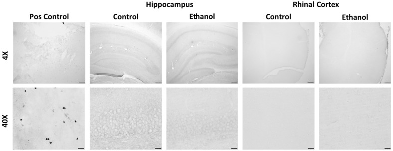
Figure 6.
Representative ED-1/CD68 images at 4X and 40X. Positive control staining is from a stroke model rat in the primary motor cortex. In our control and ethanol-treated rats there was no positive ED-1 staining throughout the brain. Shown here are the primary regions of interest: the hippocampus and the entorhinal cortex, at the T7 time point. Scale bars: top row 200 µm, bottom row 20 µm.

