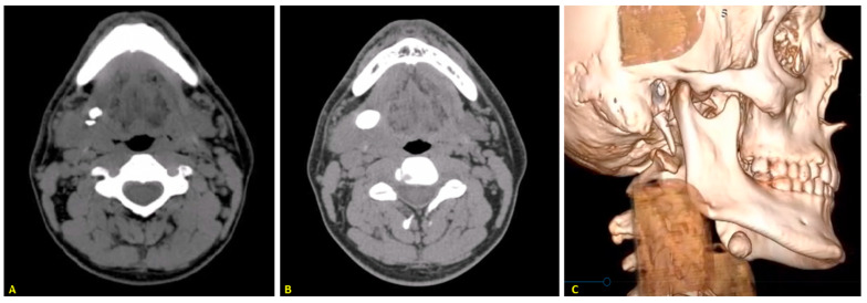
Figure 3.
(A) CT scan shows the primary stone in the hilar region and a second, smaller stone located behind it. (B) CT showing the recurrent stone in the same patient after treatment using the slitting and marsupialisation approach. (C) Three-dimensional high-resolution computed tomography reconstruction showing the recurrent stone.

