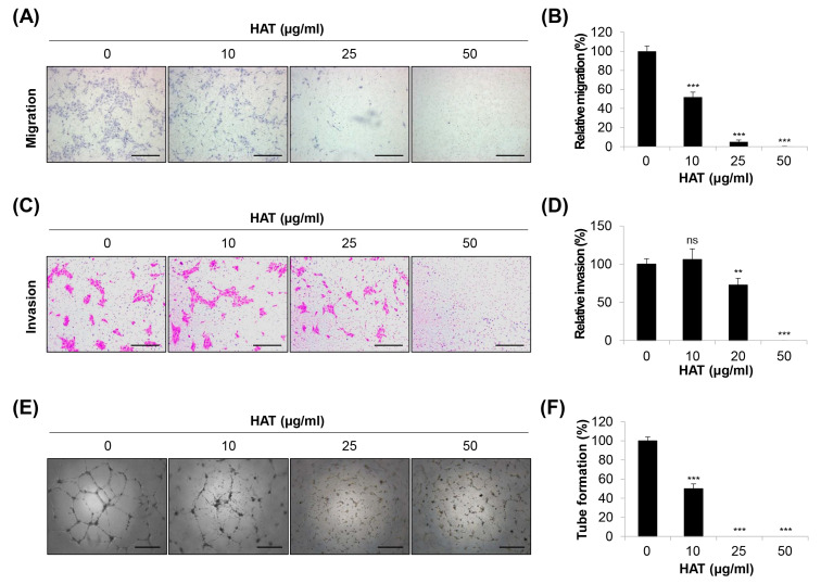
Figure 2.
Effects of HAT on the angiogenic capacity of HUVECs. The migratory ability (A,B) and invasive capacity (C,D) of HUVECs were assessed via a transwell assay after treatment with HAT. The migrated (A) or invaded (C) cells were stained and photographed (×100 magnification; scale bar = 200 μm). Relative migration (B) and invasion (D) compared to control cells were evaluated by counting the stained cells. (E) Tube formation of HUVECs after treatment with HAT was photographed (×50 magnification; scale bar = 0.4 μm). (F) Quantification of tube formation was conducted by counting the number of loops. Statistical analyses were performed using one-way ANOVA followed by Tukey’s post hoc test. ns—not significant, ** p < 0.01, *** p < 0.001 vs. control cells. HAT—hexane fraction of Adenophora triphylla var. japonica root extract; HUVEC—human umbilical vein endothelial cell; VEGF—vascular endothelial growth factor.

