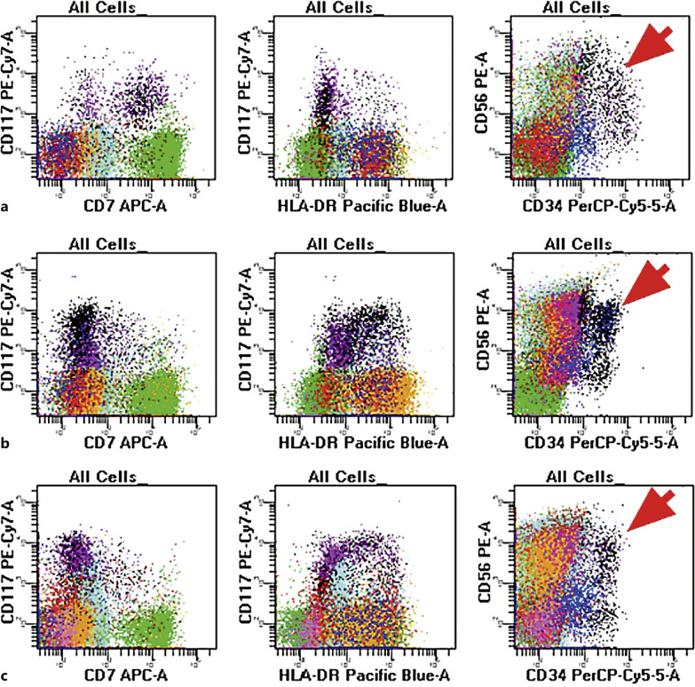
Fig. 4.
Flow cytometry evaluation of the diagnostic sample (top panel), 3 months post induction therapy (mid-panel) and 10 months post induction therapy (lower panel) in a patient with ML-DS who achieved and maintained complete remission after induction. The diagnostic sample (top panel, a) shows the presence of myeloid blasts with expression of CD7 and CD56 with the absence of HLA-DR. The post-therapy samples (mid-panel b, lower panel, c) show similar findings with persistent CD56, absent CD7, and variable HLA-DR. Aberrant CD56 expression (as indicated by the arrow) is an inherent characteristic of many DS patients and is not, in isolation, diagnostic of residual myeloid leukemia.

