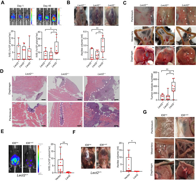
Fig. 2. Lect2−/− mice are susceptible to transcoelomic metastasis of EOC.
A The ID8/Luc cells in mice at day 0 and day 45 after inoculation. The luminescence intensity represents the tumor burden of the Lect2−/−, Lect2+/+, and Lect2+/− groups. *P < 0.05. B The ascites volume of the mice. Representative pictures of ascites taken on day 45 after the sacrifice are shown above the plot. **P < 0.01. C Quantitative plot and representative pictures of the metastatic tumors (white arrowheads) distributed in the abdominal cavity, including those on the peritoneum, mesentery, and diaphragm, among mice. **P < 0.01. D Representative H&E staining pictures of the tumor-containing areas in the diaphragm and peritoneum from mice. White dotted line: tumor/tissue interface. White arrowheads: the tumor invasive front. T: tumor, scale bar: 500 μm. E The tumor burden of Lect2+/+ mice intraperitoneally inoculated with ID8/Lect2 or the controlled ID8/Vector cells for 10 weeks. Representative luminescence photos and quantitative plots are shown. **P < 0.01. F The ascites volume of the Lect2+/+ mice inoculated with ID8/Lect2 and the control ID8/Vector cells. *P < 0.05. G Representative pictures of tumors (white arrowheads) in the peritoneum, mesentery, and diaphragm of the Lect2+/+ mice inoculated with ID8/Lect2 and the controlled ID8/Vector cells.

