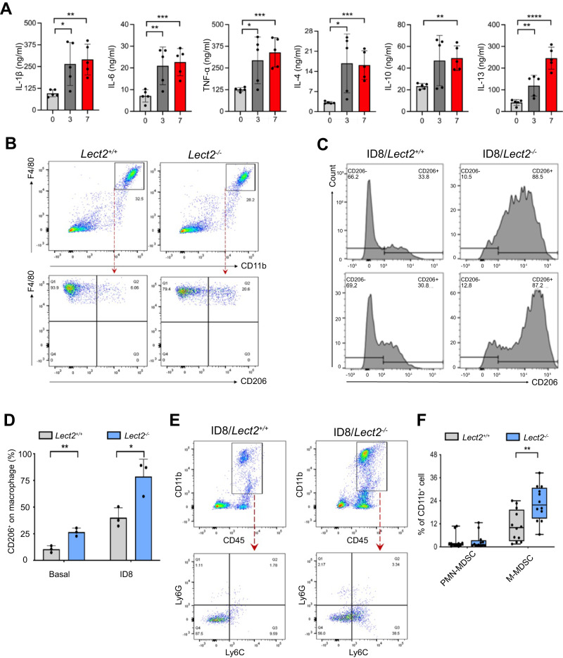
Fig. 5. Loss of Lect2 increases tumor-promoting inflammation.
A The levels of immunosuppressive (IL-4, IL-10, and IL-13) and pro-inflammatory (IL-6, IL-1β, and TNF-α) cytokines detected at weeks 0, 3, and 7 during EOC progression in the same ID8 intraperitoneal inoculation model as shown in Fig. 1C. *P < 0.05, **P < 0.01, ***P < 0.001, and ****P < 0.0001. B The representative density plots from flow cytometry analysis of peritoneal cells harvested by peritoneal lavage indicate the proportion of CD11b+F4/80+CD206+ cells in Lect2−/− and Lect2+/+ mice. C Accumulation of CD11b+F4/80+CD206+ cells in the malignant ascites of ID8/Lect2−/− and ID8/Lect2+/+ mice. Peritoneal cells were harvested 5 weeks after ID8 inoculation and analyzed by flow cytometry. D The bar chart of CD206+ macrophage proportions with or without ID8 tumor challenge in Lect2−/− and Lect2+/+ mice. *P < 0.05 and **P < 0.01. E The representative density plots from flow cytometry analysis gated on the abundance of CD45+CD11b+Ly6C+Ly6G- (M-MDSC) and CD45+CD11b+Ly6C-Ly6G+ cells (PMN-MDSC) in ID8/Lect2−/− and ID8/Lect2+/+ mice. F Quantification of the ascitic M-MDSC and PMN-MDSC in ID8/Lect2−/− and ID8/Lect2+/+ mice. **P < 0.01.

