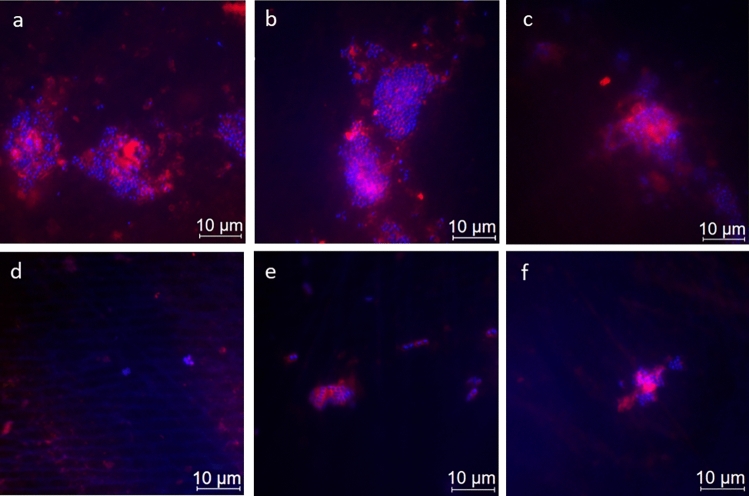
Figure 3.
Representative fluorescence microscopic images showing combined DAPI and ConA staining after 8 h of in situ pellicle formation. DAPI (blue) highlights the number of bacterial cells adhering to the enamel surface, while ConA (violet) labels bacterial glucan agglomerates. The control group (a) shows the highest number of bacteria, similar to SF rinsing (b) and SMFP treatment (c) with sporadic glucan rings and diffuse extracellular polysaccharides around almost all adherent bacteria. SnF2 (d), SnCl2 (e), and AF (f) show reduced bacterial presence with less glucan rings and extracellular polysaccharides.

