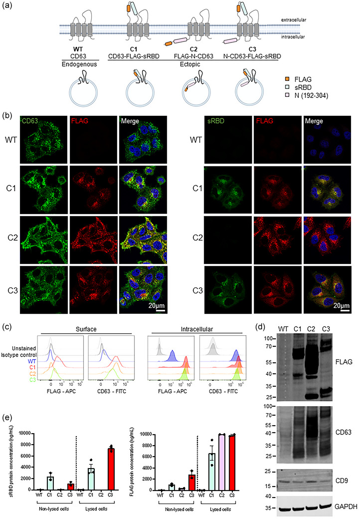
FIGURE 1.

Design of CD63‐based constructs and cell line verification. (a) A schematic representation of CD63 fusion proteins. Three CD63 fusion constructs were designed; one containing sRBD (residues 319–541 of SARS‐CoV‐2 Spike) within EC1 of CD63 (outside of sEVs), one comprising an antigenic fragment of nucleocapsid (N) protein (residues 192–304) at the N‐terminus of CD63 (inside of sEVs), and one containing both sRBD (outside) and N regions (inside). These were designated C1, C2, C3, respectively. A FLAG epitope was incorporated alongside the fusion constructs. (b) Confocal immunofluorescence of WT (mock), C1, C2 or C3 stable cells stained with anti‐CD63 (green), anti‐FLAG (red) and DAPI (blue), or anti‐sRBD (S‐Ab401.1) (green), anti‐FLAG (red) and DAPI (blue). Scale bars = 20 μm. Representative images from at least three independent experiments. (c) Surface (non‐permeabilised) and intracellular (permeabilised) flow cytometry analysis was performed on WT (mock), C1, C2 or C3 stable cell lines using antibodies against CD63 and FLAG. (d) Western blot analysis of lysates from WT (mock), C1, C2 and C3 cells. Blots were probed with anti‐FLAG, anti‐CD63, anti‐CD9 and anti‐GAPDH (loading control) antibodies. Representative images from three independent experiments. (e) FLAG and sRBD enzyme‐linked immunosorbent assays (ELISA) were performed on non‐lysed or lysed WT (mock), C1, C2 and C3 cells. n = ≥2; mean ± SEM.
