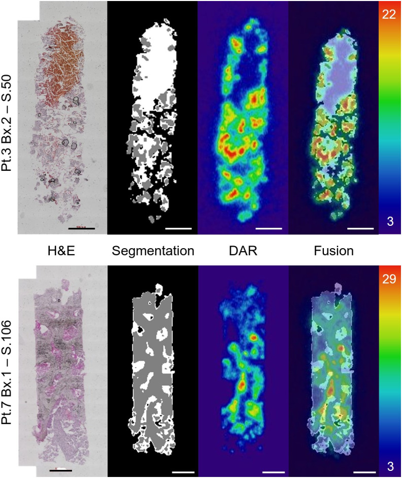
FIGURE 3.

Representative workflow of 1 section from patient (Pt.) 3 (top) and Pt. 7 (bottom). From left to right, H&E acquisition; segmented compartments of soft tissue (white), bone (gray), and background (black); registered DAR; and fused result with autoradiography of sections matching. Scale is in digital light units × 104. Training, segmentation, and autoregistration workflow schema are explained in Supplemental Figure 1. Bx. = biopsy; S. = section.
