
Figure 2. R2-only mice exhibit severely attenuated α-globin expression and are non-viable.
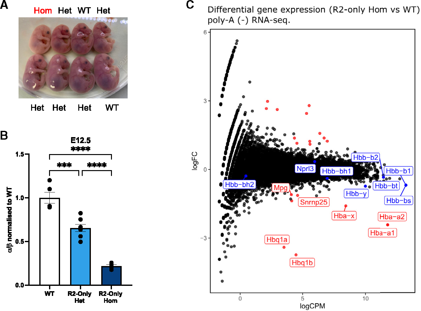
(A) Representative image of R2-only litter from heterozygote crossings. Pregnant female was sacrificed at embryonic day E17.5 and fetuses extracted. Hom, homozygotes; Het, heterozygotes.
(B) RT-qPCR comparing α-globin expression in FL erythroid cells from WT, R2-only heterozygous, and R2-only homozygous E12.5 littermates. Expression normalized to β-globin and displayed as a proportion of WT expression. Dots, biological replicates; error bars, Standard Error. Statistical analysis was performed using one-way ANOVA with a Tukey post-hoc test: ***p ≤ 0.0001, ****p ≤ 0.00001.
(C) Poly-A minus RNA-seq comparing gene expression in FL erythroid cells from WT (n = 2) and R2-only homozygous (n = 3) littermates. Red dots, differentially expressed (>2-fold); blue dots, informative erythroid genes (non-differentially expressed); black dots, no statistically significant change in expression (<2-fold). As well as reductions in transcription of the α-like globins, expression of Snrnp25 and Mpg (two genes lying upstream of the 5′ boundary of the α-globin sub-TAD) was reduced.
See also Figures S2 and S3 and Table S1.