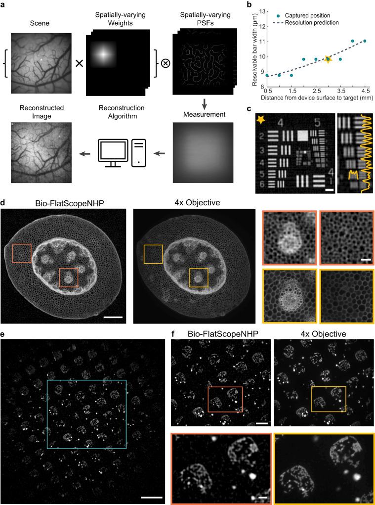
Fig. 2. High resolution images of fixed fluorescent samples compare with ground truth.
a Overview of Bio-FlatScopeNHP imaging procedure. PSFs: point spread functions. b System resolution tested using a USAF resolution target at different imaging depths. Yellow star indicates the actual imaging depth for in vivo imaging. c A representative USAF target reconstruction at the actual imaging depth for in vivo imaging. Scale bar, 100 μm. d High resolution images of a stained slice of Convallaria Rhizome captured by Bio-FlatScopeNHP and the ground truth 4X objective. Scale bars: whole Convallaria Rhizome, 500 μm; zoom-ins, 20 μm. e Patterned live spiking HEK293 cells imaged by Bio-FlatScopeNHP with integrated illumination. Scale bar, 1 mm. f Zoom-in of the blue square area in panel f of Bio-FlatScopeNHP which matched the FOV of the ground truth 4X objective. Scale bar, 500 μm. Zoom-ins below show that Bio-FlatScopeNHP is able to resolve HEK cell structures inside the square pattern. Scale bar, 100 μm.

