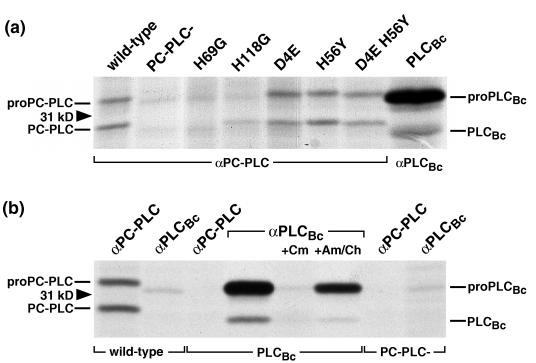
FIG. 2.
Detection of PC-PLC and PLCBc from lysates of infected cells. J774 cells were infected with strain 10403S and isogenic mutants. At 4 h after infection, cells were pulse-labeled for 30 min with [35S]methionine. Immediately after the pulse, the cells were lysed and PC-PLC or PLCBc was immunoprecipitated with the respective affinity-purified antibodies, followed by fractionation by SDS-PAGE and detection of radiolabeled proteins by fluorography. (a) Expressed phospholipase phenotypes are indicated above the lanes; positions of the 31-kDa protein size marker and pro and mature forms of the enzymes are marked on the sides. (b) Affinity-purified antibodies used for immunoprecipitation and expressed phospholipase phenotypes are shown at the top and bottom, respectively. Where indicated, prokaryotic (chloramphenicol [Cm]) and eukaryotic (anisomycin/cycloheximide [Am/Ch]) protein synthesis inhibitors were present during labeling.

