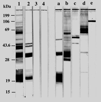
FIG. 4.
Western blot analysis of T. gondii antigens recognized by serum IgG antibodies and MAbs. Mice were injected with TSo-pulsed DC (lane 1), TSo alone (lane 2), or unpulsed DC (lane 3) or were untreated (lane 4). The following MAbs were used: 3G11 (anti-p22 [SAG2]) (lane a), 1E5 (anti-p30 [SAG1]) (lane b), 1F7 (anti-gp60 [MIC1]) (lane c), 4A7 (anti-55- and 60-kDa [ROP2 to ROP4]) (lane d), and 4A11 (anti-p100 [MIC2]) (lane e). The molecular masses (in kilodaltons) of protein standards are given on the left.

