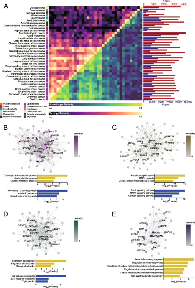
Fig. 5.
Comparative analysis of interactome networks specific to malignant cells across different tumors. A The heatmap depicts the comparison of interactome networks specific to malignant cells identified from different tumor types. The red gradient panel represents the topology similarity estimated from shared nodes’ topological specificity. The green gradient panel represents the edge similarity estimated from shared edges’ interaction strengths. Network sizes are shown by number of nodes (red bars) and number of edges (blue bars). The corresponding tumor types and physiological cell types of different malignant cells are also labeled. B–E The graph plots depict four representative core subnetworks identified from the shared network of gastric cancer, colorectal cancer, and pancreatic ductal adenocarcinoma. The centrality of each gene implicated in the subnetwork is labeled in color. The bar plots under each graph plot shows the GO (yellow bars) and KEGG (blue bars) pathway enriched for each core subnetwork

