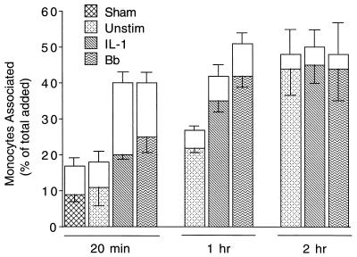
FIG. 1.
Time course of the migration of monocytes across HUVEC stimulated with B. burgdorferi or IL-1. Monocytes were incubated for indicated times with HUVEC-amnion cultures that had been pretreated for 8 h with either control medium (Unstim), a sham preparation, 5 U of IL-1 per ml, or B. burgdorferi (Bb) at a ratio of 10 Bb/EC. Transendothelial migration was assessed as described in Materials and Methods. The total height of each bar represents the number of monocytes associated with each culture as a percentage of the total number added. The lower (patterned) portion of each bar represents the percentage that migrated beneath the endothelium; the upper (unfilled) portion represents the percentage adherent to the apical surface. Bars represent the means ± SD of three to four replicate samples. This experiment was repeated twice with similar results.

