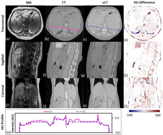
FIGURE 5.

Traverse, sagittal, and coronal images of a representative patient. MRI, CT, and sCT images and the HU difference map between CT and sCT are presented. The CT (solid line) and sCT (dashed line) voxel‐based HU profiles of the traverse images are compared in the lowermost panel. Reprinted by permission from British Journal of Radiology, MRI‐based treatment planning for liver stereotactic body radiotherapy: validation of a deep learning‐based synthetic CT generation method by Liu et al.108© 2019.
