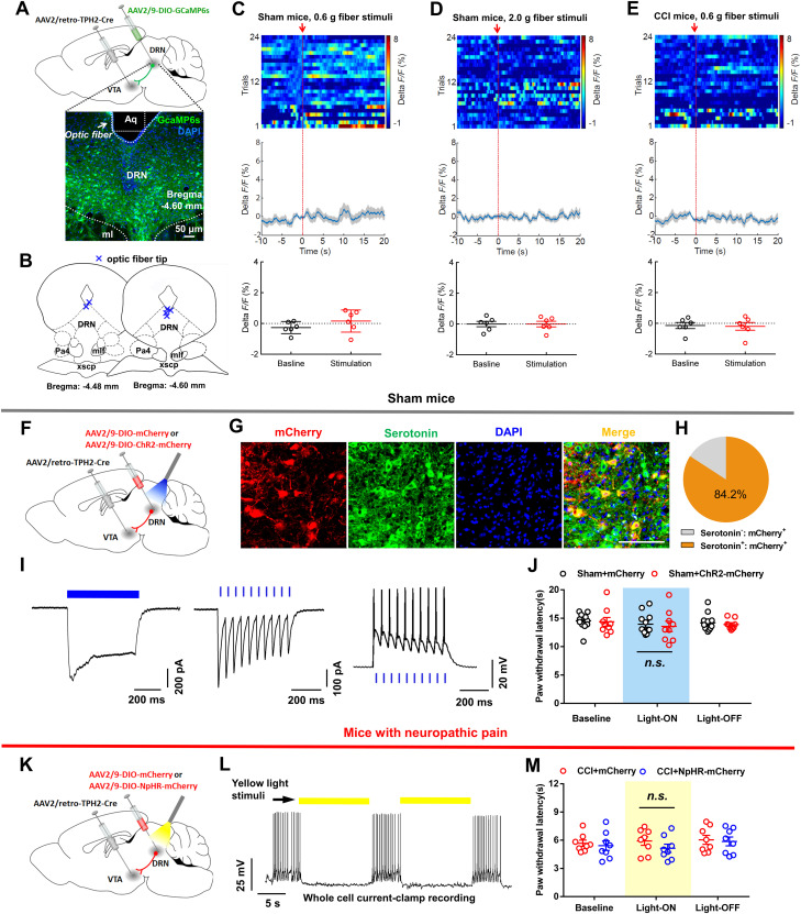
Figure 6.
DRN-VTA serotonergic input plays insignificant roles in pain regulation. A, Experimental design for expression of GCaMP6s in DRN-VTA serotonin neurons and calcium imaging in freely moving mice. Scale, 50 μm. B, The position of optic fiber tips (blue cross) in the DRN of mice used for photometry recording. C, Response of DRN-VTA serotonin neurons following mechanical stimulation (von Frey, 0.6 g) in sham-operated mice (top, activity heatmap where each line corresponds to a single stimulation event; middle, average activity trace of 24 stimulation events; bottom, quantification of change in fluorescence in mice following exposure to stimulation). The red arrow indicates time point at which stimulation occurred (paired t test, t5 = 1.141, p = 0.3055, n = 6 mice). D, Response of DRN-VTA serotonin neurons following painful stimulation (von Frey, 2.0 g) in sham-operated mice. The arrow indicates time point at which stimulation occurred (paired t test, t5 = 0.0164, p = 0.9876, n = 6 mice). E, Response of DRN-VTA serotonin neurons following mechanical stimulation (von Frey, 0.6 g) in CCI mice. The arrow indicates time point at which stimulation occurred (paired t test, t5 = 0.3216, p = 0.7608, n = 6 mice). F, Schematic showing viral injection into the DRN and VTA. G, H, Confocal images (G) and quantification (H) showed that the majority of mCherry-expressing DRN cells were serotonin-positive (n = 5 sections from 3 mice). I, In ChR2-mCherry infected DRN neurons, long duration (500 ms, left panel) or short bursts (20 Hz, 5 ms, middle panel) of blue light induced temporally precise inward photocurrents and short bursts (20 Hz, right panel) of blue light produced spikes. J, There was no difference in thermal (left) and mechanical (right) pain thresholds between mCherry-expressed and ChR2-mCherry-expressed sham mice when they received blue light illumination (two-way repeated measures ANOVA, group × epoch interaction, left: F1, 19 = 0.03038, p = 0.8635; right: F1, 19 = 0.5873, p = 0.4529; n = 9, 12 mice/group). K, Schematic showing viral expression of mCherry or NpHR-mCherry in DRN-VTA serotonin neurons. L, The whole-cell recording revealed that yellow light illumination (15 s) of an NpHR-mCherry-positive DRN neuron reliably inhibits its firing activity. M, Optic inhibition of DRN-VTA serotonin neurons could not change the thermal hyperalgesia (left) and mechanical allodynia (right) in CCI mice (two-way repeated measures ANOVA, group × epoch interaction, left: F1, 14 = 0.3204, p = 0.5803; right: F1, 14 = 0.8597, p = 0.3695; n = 8 mice/group). Data are represented as mean ± SEM.

