Figure 1.
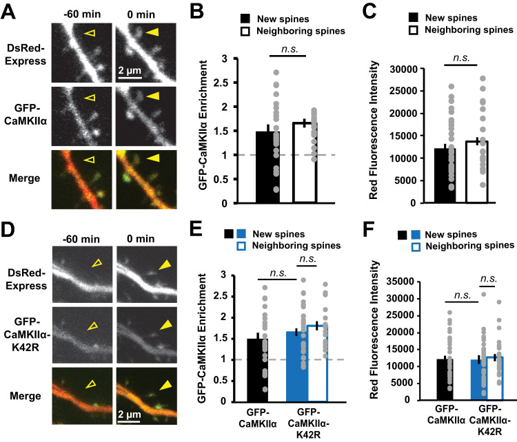
GFP-CaMKIIα enrichment in new spines is comparable to that in size-matched neighboring spines. A, Images of a dendrite from a hippocampal CA1 neurons in slice culture (DIVs 7–9) expressing DsRed-Express (red) and GFP-CaMKIIα (green) before (open arrowhead) and after (filled arrowhead) spontaneous new spine outgrowth. B, Enrichment (spine:dendrite ratio) of GFP-CaMKIIα in new spines (n = 33 spines/16 cells) was comparable to that in size-matched neighboring spines (n = 21 spines/16 cells). C, Neighboring spines used for enrichment calculations in B were size-matched to new spines (p = 0.62). D, Images of dendrites from CA1 neurons expressing GFP-CaMKIIα-K42R (green) and DsRed-Express (red) before (open arrowhead) and after (filled arrowhead) spontaneous new spine outgrowth. E, Enrichment of GFP-CaMKIIα-K42R in new spines (n = 39 spines/14 cells) was comparable to that in size-matched mature neighboring spines (n = 39 spines/14 cells). Importantly, no difference in relative enrichment was found between new (filled bars) or size-matched neighboring spines (open bars) in the WT (black) and K42R (blue) conditions (new, p = 0.4; neighbors, p = 0.99). Data for GFP-CaMKIIα-WT new spine enrichment is from B. F, Neighboring spines used for enrichment calculations in E were size-matched to new spines (p = 0.99). No difference in new spine size was found between WT (black) and K42R (blue; p = 0.99). Data for GFP-CaMKIIα new spine size is from C. Two-way ANOVA with Bonferroni’s multiple-comparisons test.

