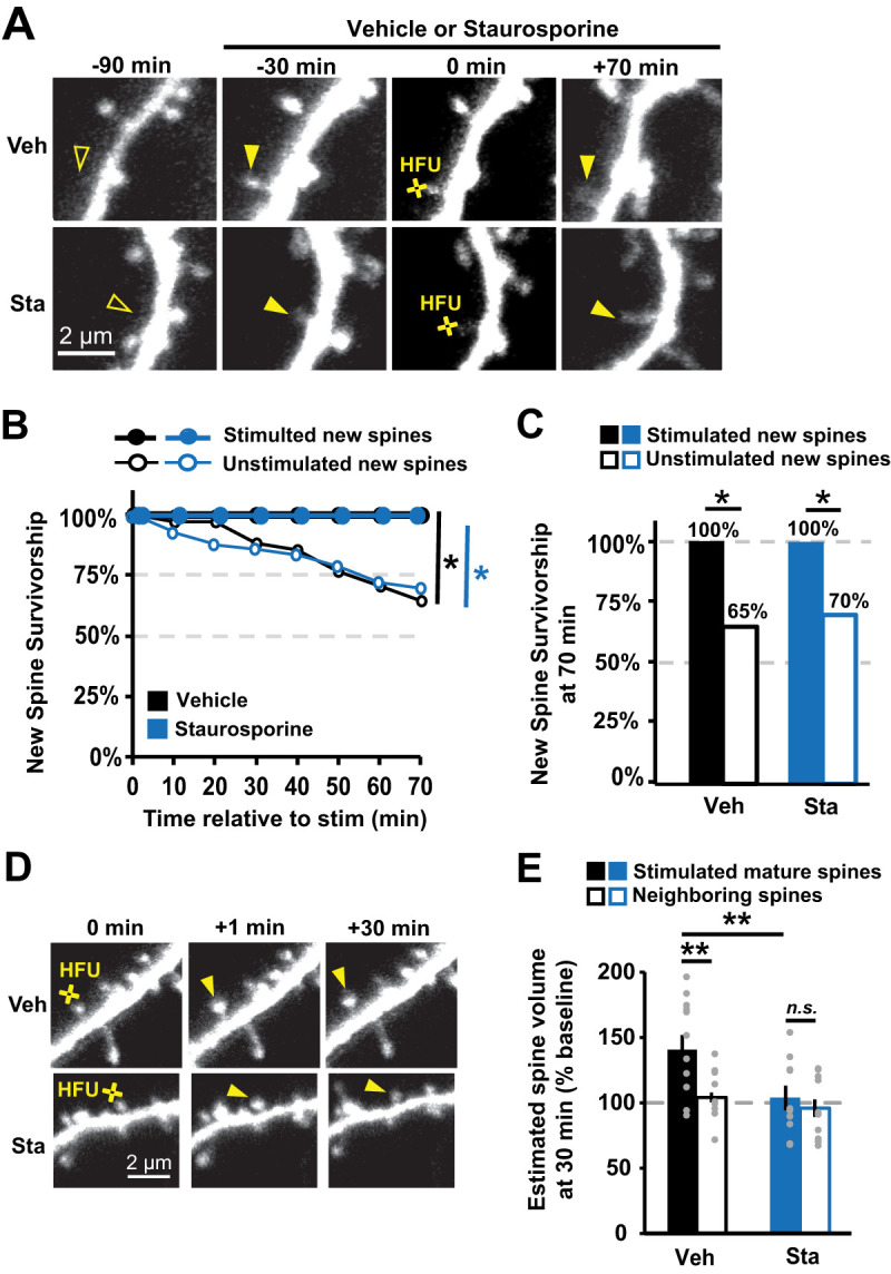
Figure 3.

Inhibition of CaMKIIα kinase activity with staurosporine does not impair activity-dependent new spine stabilization. A, Images of green fluorescence showing dendrites from GFP-transfected CA1 neurons in slice culture (DIVs 7–9). One new spine (filled arrowhead at 0 min) per neuron was stimulated with HFU at 0 min, following 30 min pre-incubation in either vehicle (Veh, top row) or 1 µm staurosporine (Sta, bottom row). B, Survivorship of stimulated new spines (filled circles; Veh, 9 spines/9 cells; Sta, 11 spines/11 cells) was enhanced relative to unstimulated new spines on the same cells (open circles; Veh, 33 spines/9 cells; Sta, 43 spines/11 cells) in both vehicle (black) and staurosporine (blue) conditions. C, Survivorship of HFU-stimulated new spines (filled bars) at 70 min was increased compared with unstimulated new spines (open bars) on the same cells in both vehicle (black) and staurosporine (blue) conditions. D, Images of green fluorescence showing dendrites on GFP-transfected CA1 neurons before and after HFU (yellow circle). E, Incubation with 1 µM staurosporine (Sta; filled blue; n = 10 spines/10 cells) blocked HFU-induced long-term growth of mature spines (101 ± 9%; p = 0.03), which was intact in vehicle conditions (Veh; filled black; n = 12 spines/12 cells; 140 ± 11%; p = 0.99). Volume of unstimulated neighbors was unchanged (open bars; Veh, 104 ± 3%; p = 0.99; K42R, 96 ± 7%; p = 0.99). Log-rank task in B, Barnard's exact test in C, and two-way ANOVA with Bonferroni’s multiple-comparisons test in E. *p < 0.05, **p < 0.01, ***p < 0.001.
