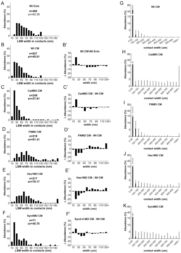
Fig 4. LSM widths in contacts.
(A–F) Width frequency distributions in ectoderm (A) for comparison, in CM (B) and in various CM morphants (C–F). n, number of width measurements from 18, 4, 6, 8, 3 TEM images, respectively; av., average. (B’–F’) Corresponding difference (ΔAbundance) spectra comparing width distributions of CM to ectoderm (B’), and of morphants to normal CM (C’–F’). (G–K) Comparison of widths of LSM-containing (black parts of bars) and LSM-free (grey parts of bars) contacts, using the data from (B–F) and S1 Fig.

