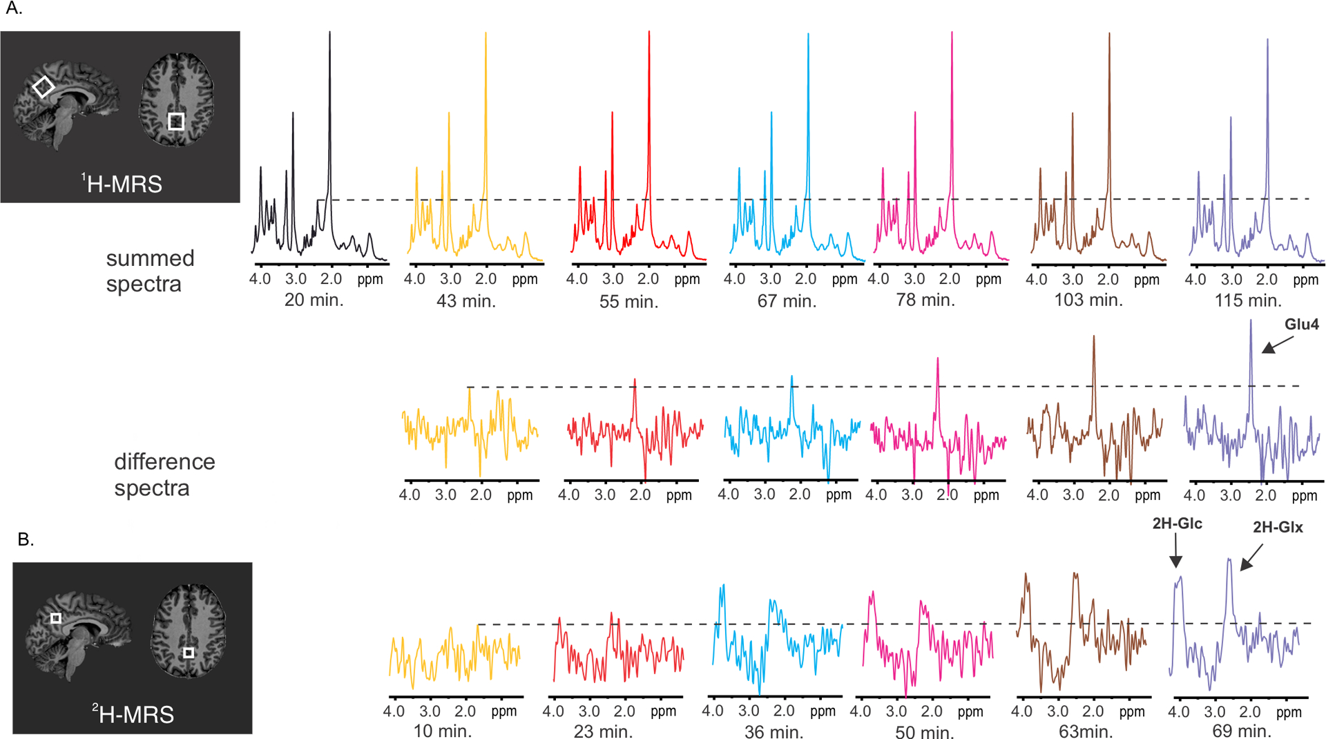
Fig. 2 |. Example of MR spectra obtained from one voxel in the posterior cingulum with single voxel 1H-MRS (panel A) and with 2H-MRSI (panel B) in one participant at 7 Tesla.

The displayed spectra were obtained after peroral administration of 2H-Glc. A. The summed spectra show decreasing signal at the resonance frequency of -C41H2- in the glutamate molecule (2.34 ppm) due to enrichment of the glutamate pool with 2H (either -C42H2H- or -C42H1H). The robust change at the 4th carbon position is documented by the increasing signal amplitude at 2.34 ppm in the difference spectra calculated by subtraction of the respective spectrum from the last session minus the first session. B. 2H-MR spectra reflect an increase in the deuterated fraction of Glc and Glx (Glu+Gln). The insets with anatomical images show location of voxels in the posterior cingulate gyrus. The voxel dimensions were 22×20×20 mm for single-voxel proton MRS, whereas voxel selected from 2H-MRSI data had volume of (12.5 mm)3.
