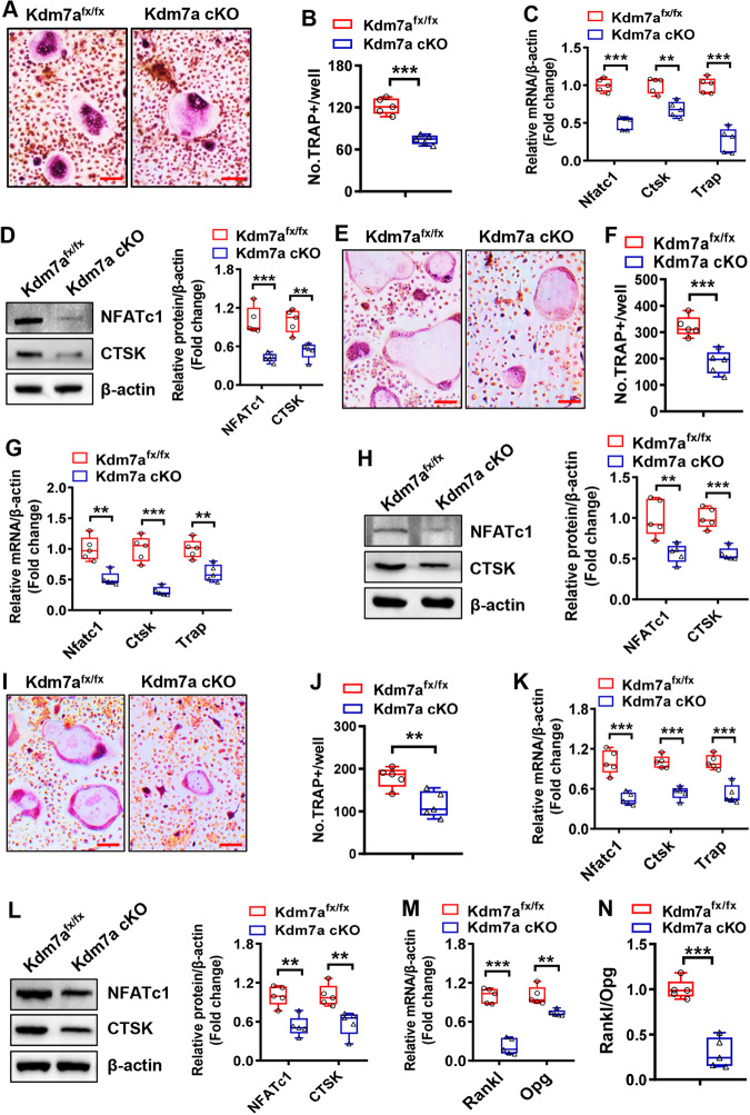
Fig. 5. Kdm7a deletion in osteoprogenitor cells suppressed osteoclast formation from osteoclast precursor cells.
Bone marrow cells were isolated from 6-week-old Kdm7a cKO and Kdm7afx/fx mice and induced to allow osteoclast differentiation. Eight days after induction, TRAP staining was performed (A) and the number of osteoclasts was counted (B). The mRNA (C) and protein (D) expression levels of osteoclast-specific genes were measured 5 days after induction using RT-qPCR and Western blotting, respectively. Wild-type BMM cells were co-cultured with BMSCs derived from Kdm7a cKO or Kdm7afx/fx mice in cell-cell contact manner (E–H) or in transwell manner (I–L). The co-cultures were induced to allow osteoclast differentiation. Eight days after induction, TRAP staining was performed (E, I) and osteoclast number (F, J) was counted. The mRNA (G, K) and protein (H, L) expression levels of osteoclast-specific genes were measured 5 days after induction using RT-qPCR and Western blotting, respectively. The mRNA levels of Rankl and Opg in BMSCs were examined (M) and the Rankl/Opg ratio was calculated (N). Image scale in (A, E, I): 100 μm. Data are presented as box-and-whiskers plots, n = 5. Comparisons were conducted using Student’s t test. **p < 0.01, ***p < 0.001.

