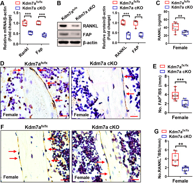
Fig. 6. Kdm7a deletion in osteoprogenitor cells downregulated FAP and RANKL expression in female mice.
Long bone BMSCs were isolated from Kdm7a cKO and Kdm7afx/fx mice. The mRNA and protein levels of RANKL and FAP were examined by RT-qPCR and Western blotting, respectively (A, B). Serum RANKL levels were measured in 24-week-old female mice using ELISA (C). IHC staining of FAP (D) and RANKL (F) was performed. The numbers of FAP-positive (E) and RANKL-positive (G) cells on trabeculae were counted. Data are presented as box-and-whiskers plots, n = 5 in (A, B); n = 10 in (C); n = 9 in (E, G). Comparisons were conducted using Student’s t test. **p < 0.01, ***p < 0.001.

