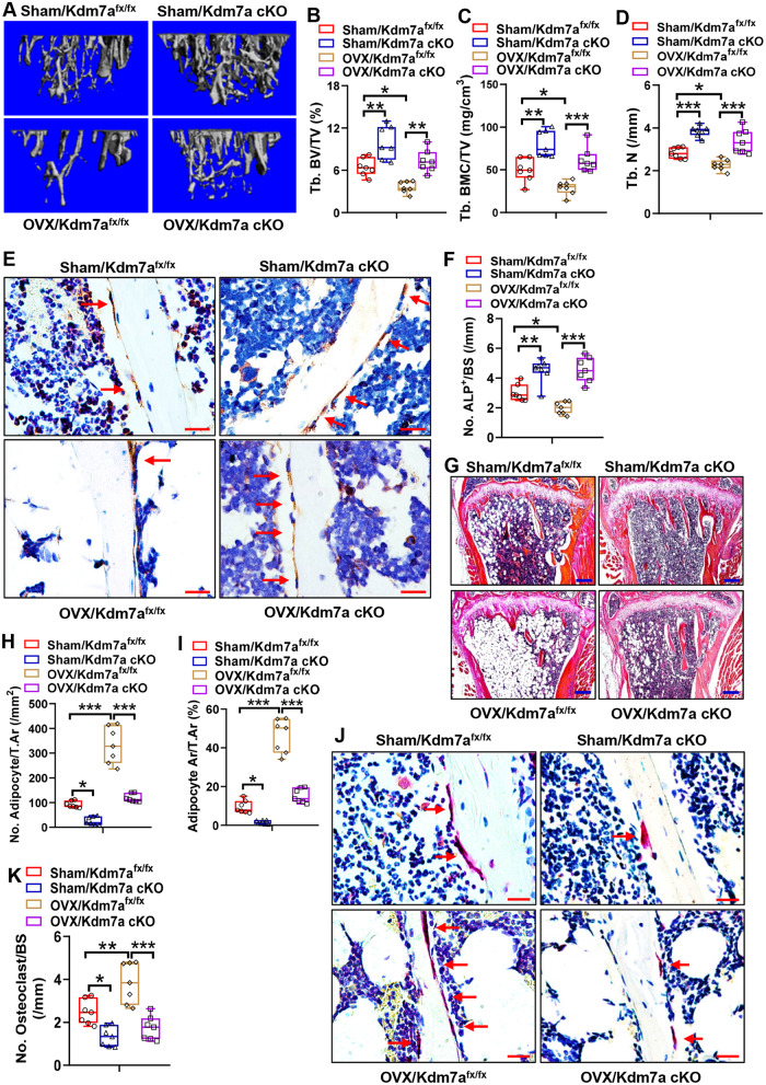
Fig. 9. Deletion of Kdm7a in osteoprogenitor cells prevented bone loss in OVX mice.
μCT analyses of tibial metaphyseal bone mass were performed and the reconstruction images are shown (A). Histomorphometric parameters including Tb. BV/TV, Tb. BMC/TV and Tb. N were measured (B–D). ALP IHC staining was performed (arrows indicate osteoblasts) (E). Image scale: 20 μm. The number of trabecular osteoblasts in the tibial metaphysis was counted (F). H&E staining was performed (G). Image scale: 200 μm. The number and area of adipocytes in marrow were quantified (H, I). TRAP staining was performed (J). Image scale: 20 μm. The number of osteoclast was counted (K). Data are presented as box-and-whiskers plots, n = 7. Comparisons were conducted using two-way ANOVA followed by Tukey’s test. *p < 0.05, **p < 0.01, ***p < 0.001.

