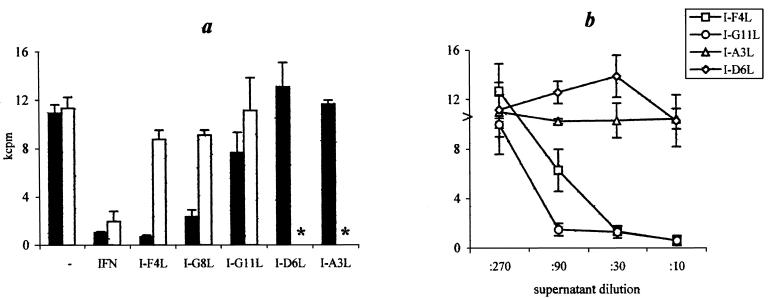
FIG. 5.
Stimulation of macrophage antimycobacterial activity by lung T-cell clones and their supernatants. Peritoneal macrophages (6 × 10⁴/well) were loaded with 12 × 104 live mycobacteria, and either 6 × 10⁴ T-cell clones (a) or serial dilutions of their cultural supernatants (b) (see the footnote to Table 2) were added to cultures. Following 96 h of incubation, activity of mycobacterial growth was measured as [3H]uracil uptake (mean ± standard deviation). (a) Solid bars, syngeneic I/St system; open bars, allogeneic system (B6 macrophages cocultured with I/St T cells); asterisk, not tested. (b) Arrowhead, no T-cell clone supernatant was added. The results of one of three representative experiments are shown.

