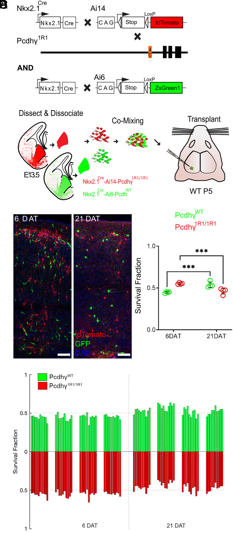
Fig. 3.
Most MGE-derived cINs survive the deletion of most Pcdhγ, except for PcdhγC4. (A) Diagram of genetic crosses between MGE/POA-specific reporter Nkx2.1Cre;Ai14 and Pcdhγ1R1 mice. Control cells were derived from Nkx2.1Cre;Ai6 mice while Pcdhγ1R1 mutant cells were derived from Nkx2.1Cre;Ai14;Pcdhγ1R1/1R1 mice. (B) Diagram of the transplantation protocol. The MGEs from E13.5 Pcdhγ1R1 homozygous or control embryos were dissected, dissociated, and mixed in similar proportions. The mixture of GFP+ (PcdhγWT) and tdTomato+ (Pcdhγ1R1/1R1) cells was grafted into the cortex of WT neonate mice. (C) Left—Confocal images from the cortex at 6 and 21 DAT. The transplanted cells are labeled with GFP (PcdhγWT) or tdTomato (Pcdhγ1R1/1R1). Right—Quantifications of the transplanted cells that survived at 6 and 21 DAT. Note that both the GFP and tdTomato labeled cells underwent PCD between 6 and 21 DAT, but both cell types survived to similar levels. (D) Survival fraction quantifications from (C) shown by individual brain sections (each bar) and separated by animals at 6 and 21 DAT. Scale bar = 50 μm, nested ANOVA, ***P = 0.0026, n = 4 mice per time point and 10 brain sections per mouse. DAT 6 cell counted = 11,063, DAT 21 cells counted = 7,751.

