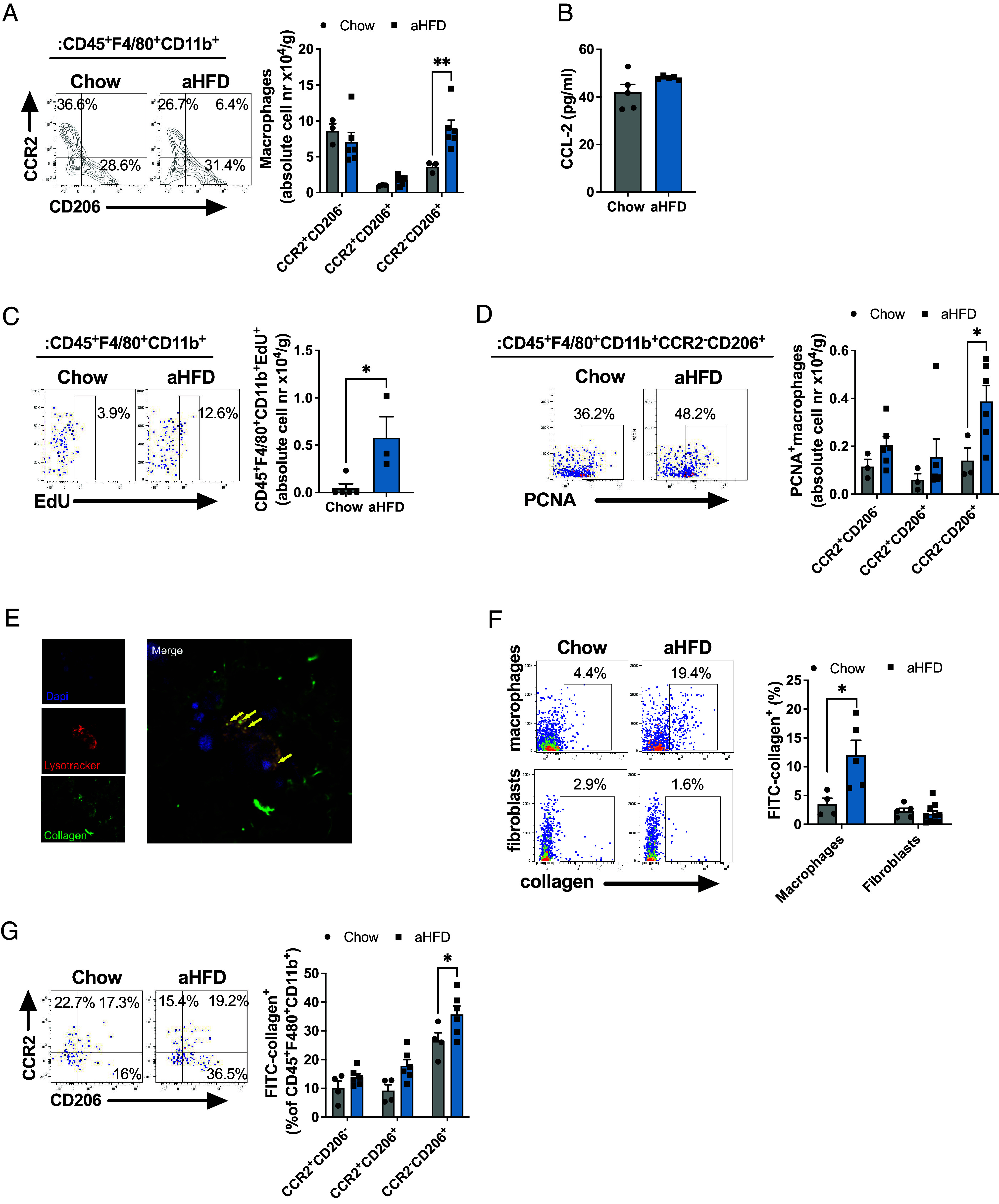
Fig. 2.

Acute HFD drives proliferation of SAT resident macrophages that engage in collagen endocytosis. (A) Representative dot plot and number of infiltrated (CCR2+CD206− and CCR2+CD206+) and resident (CCR2−CD206+) macrophages (single live CD45+F4/80+CD11b+) per gram of SAT from chow (n = 7, some samples pooled) and aHFD (n = 6) mice. (B) Serum levels of CCL-2 from chow (n = 5) and aHFD mice (n = 4). (C) Representative dot plot and number of proliferating (EdU+) macrophages (single live CD45+F4/80+CD11b+) per gram of SAT from chow (n = 5) and aHFD (n = 4) mice. (D) Representative dot plot and number of PCNA+ total (single live CD45+F4/80+CD11b+), infiltrated (CCR2+CD206− and CCR2+CD206+ of total), and resident (CCR2−CD206+ of total) macrophages per gram of SAT of chow (n = 7, some samples pooled) and aHFD (n = 7) mice. (E) Representative image of collagen-endocytosing macrophages. Ex vivo sorted macrophages (F4/80+ SVF of SAT) where immersed in neutralized FITC-collagen and left to polymerize. Collagen (green) forms the fibers similar to in vivo settings. Lysosomes were stained with Lysotracker red (red). Nuclei were stained with Dapi (blue). Macrophages endocytose collagen which can be noted as co-localization of green (collagen) and red (lysosomes) signal. (F) Collagen endocytosis assay. Frequencies of FITC-collagen+ macrophages (single live CD45+F4/80+CD11b+) and fibroblasts (single live CD45-PDGRFα+) from SAT of chow and aHFD mice (n = 10/group). Cells were magnetically sorted (macrophages: F4/80+ of SVF, fibroblasts: F4/80−CD45−CD90.2+ of SVF). (G) Collagen endocytosis assay- subset distribution of collagen endocytosing macrophages. Frequencies of FITC-collagen+ infiltrated (CCR2+CD206− and CCR2+CD206+) and resident (CCR2−CD206+) macrophages (single live CD45+F4/80+CD11b+). Data are presented as mean ± SEM and are representative of two or more independent experiments. Unpaired student’s t tests (B, C, and F), two-way ANOVA with Fisher’s post hoc test (A, D, and G). *P < 0.05, **P < 0.01
