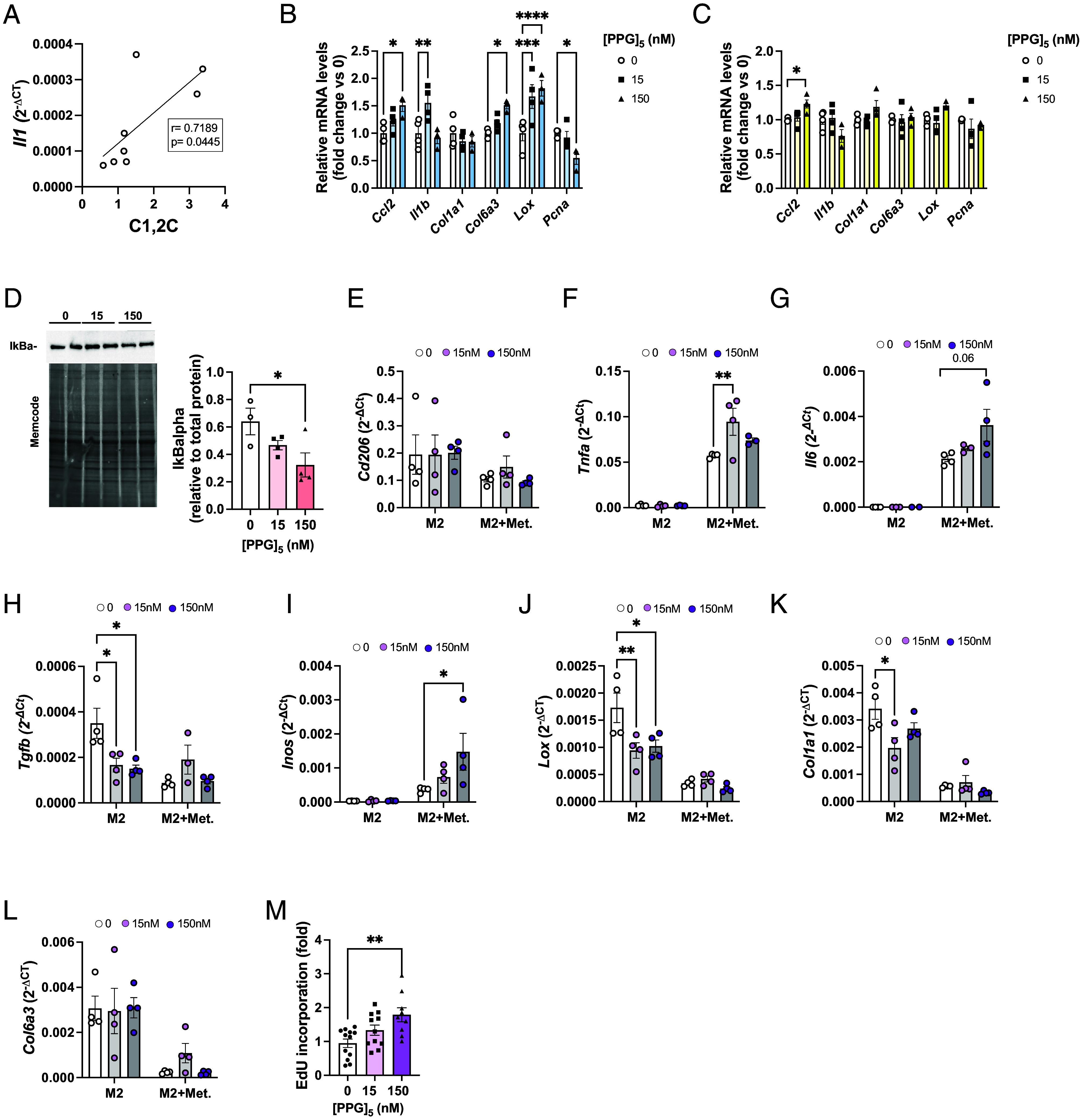
Fig. 5.

Collagen fragments induce inflammation and fibrosis in pre- and adipocytes, but proliferation in M2-like macrophages. (A) Pearson correlation of mRNA expression of Il1 and the amount of collagen fragments in the SAT of mice treated with anti-CD206 or IgG control. mRNA expression (relative to untreated cells) after in vitro treatment of (B) 3T3-L1 fibroblasts and (C) differentiated 3T3-L1 adipocytes. (D) Western blot analysis of IkBalpha in 3T3-L1 pre-adipocytes after 24 h treatment with [PPG]5. Band intensity was normalized to total protein amount on the membrane. (E–L) mRNA expression in M2 BMD macrophages after 24 h treatment with [PPG]5 (n = 3 to 4 per group) in the presence or absence of metabolic cocktail (25 mM glucose, 0.5 mM palmitate-BSA, 10 nM insulin). Expression is relative to Bactin. (M) EdU incorporation in M2 BMD macrophages after 24 h treatment with collagen mimetic peptide (15 and 150 nM, n = 9 to 12/group). Data are presented as mean ± SEM and are representative of three independent experiments (A–D). One significant outlier value was detected with Grubb’s outlier test (P < 0.05) and removed (F and G). One-way (B–D and M) and two-way (E–L) ANOVA. *P < 0.05, **P < 0.01.
