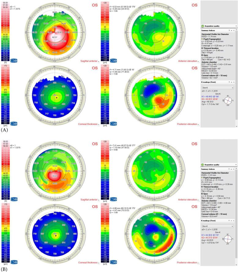
Figure 4.
(A) Preoperative and (B) postoperative corneal topographies (Sirius corneal topography device; Costruzioni Strumenti Oftalmici, Florence, Italy) of a pediatric patient with type 2 asymmetry cone (80% of the cone was on one side of steepest meridian) and Amsler – Krumeich [13] grade 2 (Kaverage 48 – 53 D)

