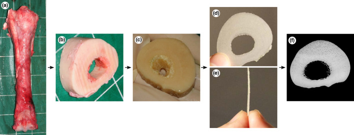
FIGURE 2.

Bone processing from post‐mortem collection to microradiograph: (a) dorsal view of the right third metacarpal bone immediately post‐mortem, (b) transverse 20 mm thick slice cut with a band‐saw, (c) slice after 5 days of immersion in bacterial pronase detergent and subsequent fixation for 7 days in 70% ethanol, (d) proximodistal and (e) lateral view of 250 μm thick sections cut with an annular saw, (f) microradiograph of 250 μm thick section. Lateral to the left, dorsal to the top (c, d, f).
