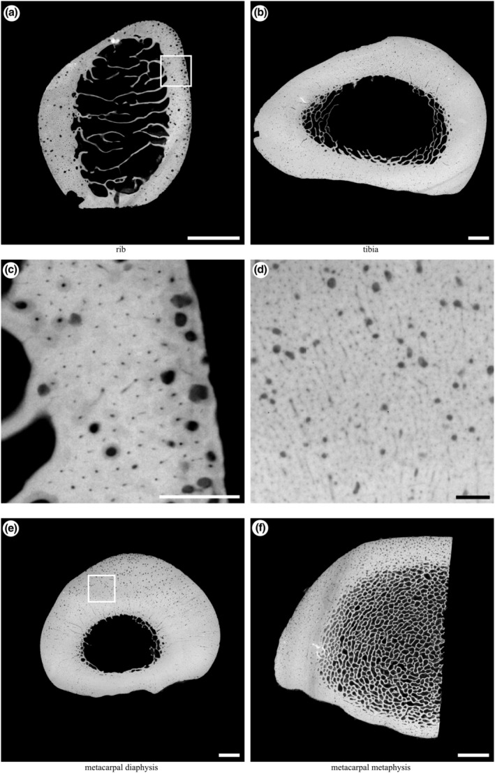
FIGURE 3.

Example microradiographs obtained from each section: (a) transverse section of the mid‐diaphysis of the left tenth rib (5× magnification); (b) transverse section of the distal third region of the right tibia (2×magnification); (e) transverse section of the mid‐diaphysis of the right third metacarpal (2× magnification); (f) transverse section of the lateral half of the distal metaphysis of the right third metacarpal (3× magnification). Box in (a) magnified in (c); box in (e) magnified in (d). Scale bars: a, b, e, f, 5 mm; c, d, 1 mm. Lateral to left, cranial/dorsal to top.
