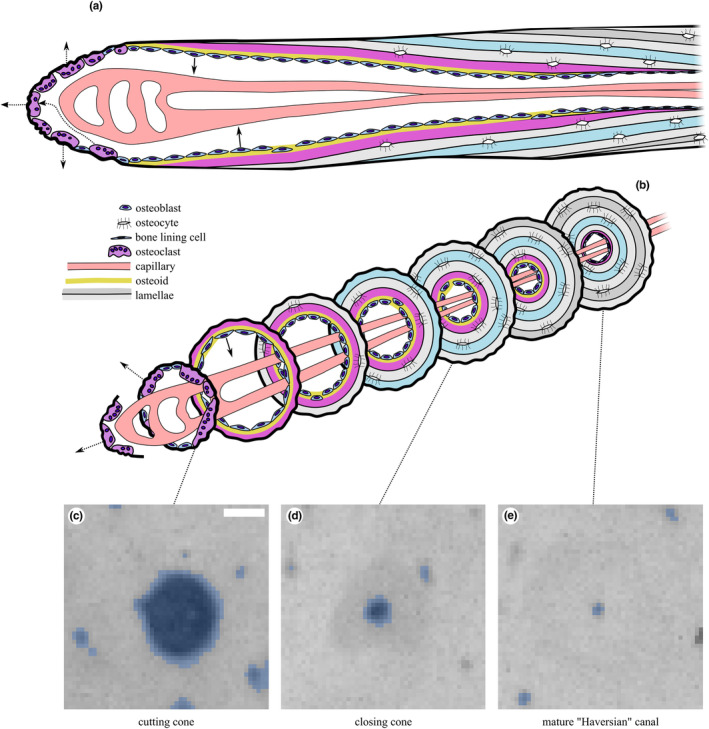
FIGURE 4.

Illustration of secondary osteonal remodelling “basic multicellular unit” in cortical bone in (a) longitudinal section and (b) a series of transverse sections along its length. Representative microradiographs of the (c) cutting cone, (d) closing cone and (e) mature Haversian canal, shown with binary masks derived from processing with Fiji's Analyze Particles overlaid in blue. In this study, we classify canals into large (Ca.Ar >0.04 mm2; (c)) and combined large and small (Ca.Ar >0.002 mm2; (c + d)) and ignore the smallest canals (Ca.Ar ≤0.002 mm2; (e)). Scale bar 100 μm. Modified from (Doube, 2022) under CC‐BY licence terms.
