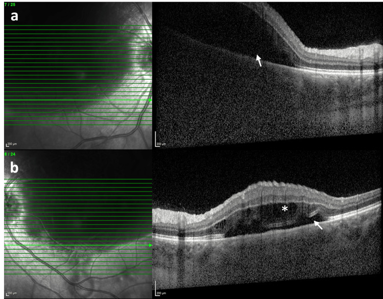
Figure 1. Spectral domain optical coherence tomography scan at first presentation.
The horizontal SD-OCT scan demonstrates marked serous retinal detachment (white arrow) in the right eye (a) and a shallow serous detachment (white arrow) with intraretinal cysts (asterisk) in the left eye (b). The extent of the lesion is partially visible in the infrared reflectance image in both eyes.
SD-OCT, Spectral domain optical coherence tomography

