
Figure 3.
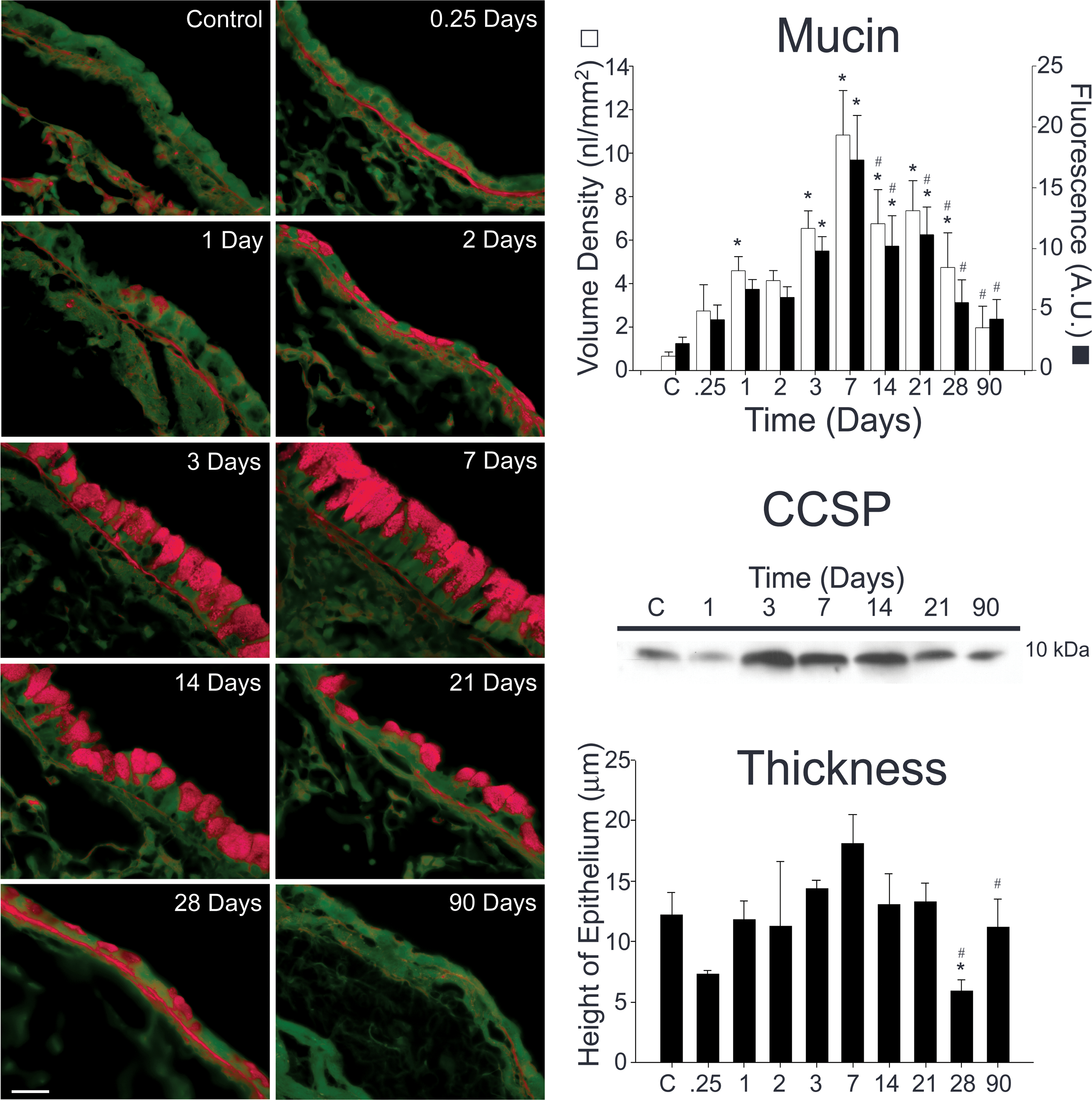
Mucin and CCSP content both increase during allergic airway inflammation. Tissue sections from antigen challenged mice were stained using a periodic acid fluorescent Schiff (PAFS) method (left panels). Mucin content increases as early as 6 h (0.25 days) following antigen challenge, is significantly increased 1 day following antigen challenge, and is maximal at 1 week (right, upper panel). Mucin content decreases significantly from peak levels 14–28 days following antigen exposure (right, upper panel). CCSP content of the lung was measured by Western blotting. CCSP is produced constitutively in the lungs of control mice (right, middle panel). The amount of CCSP increases at days 3–14 and returns to baseline at day 21. There was also an increase in the thickness of the airway epithelium that peaked at day 7 (right, lower panel). While this increase was not statistically significant, the decrease in thickness (seen coincidentally with decreasing mucin and CCSP levels) was significantly lower than unchallenged mice (day 28) and day 7 mice (days 28 & 90). Three to six animals were used for each time point except for 0.25 days (n=2). * indicates statistically significant difference from control, and # signifies statistically significant difference from day 7.