
Figure 7.
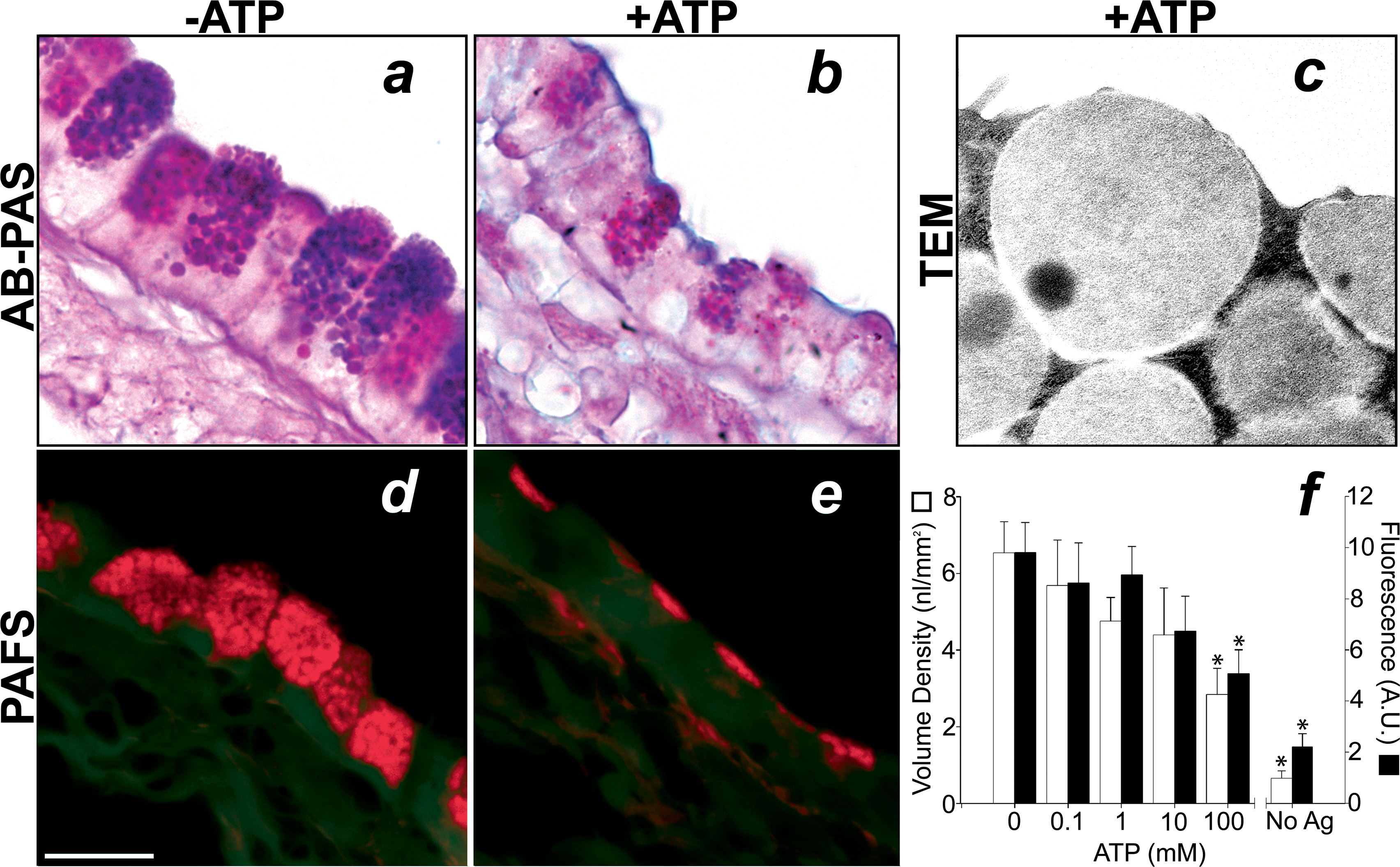
Mucin is secreted from airway epithelial cells in a ligand-regulated manner. In mice 3 days after antigen exposure, increasing concentrations of aerosolized ATP (0.1–100 mM, 5 min) cause a dose dependent decrease in AB-PAS and PAFS staining. Top. In the absence of an ATP aerosol, mucous cells in antigen challenged mice contain large granular stores of AB-PAS positive mucin (a). Exposure of mice to ATP (100 mM, 5 min) causes an acute decrease in intracellular AB-PAS positive mucin stores (b). In mice euthanized immediately after ATP inhalation secretory granule fusion events are frequently observed (c). Bottom. The degree of ATP-induced secretion was measured in PAFS stained sections from antigen challenged mice that received aerosolized ATP (d) compared to animals that received aerosolized saline alone (e). The dose dependence of ATP-stimulated mucin secretion was quantified using volume density measurements (open bars) and fluorescence quantitation (closed bars) in PAFS stained sections (f). Scale bar is 10 μm in a, b, d, and e and 250 nm in c. * indicates statistically significant difference from antigen challenged mice not treated with ATP aerosol. Data from unchallenged mice are the same as in figure 4 (note different scale).