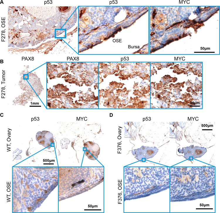
Figure 4. Molecular histology of ovarian surface epithelium.
(A) An OvTrpMyc at 16mo of age with clear ovarian surface epithelium (OSE) staining of p53 and MYC. (B) The intra-abdominal tumor extracted from the mouse shown in (a) exhibited PAX8, p53, and MYC staining. (C) A wild-type mouse at 12mo of age is negative for p53 or MYC staining in OSE. (D) Example of an OvTrpMyc mouse with negative OSE staining of p53 and MYC, at 12mo of age.

