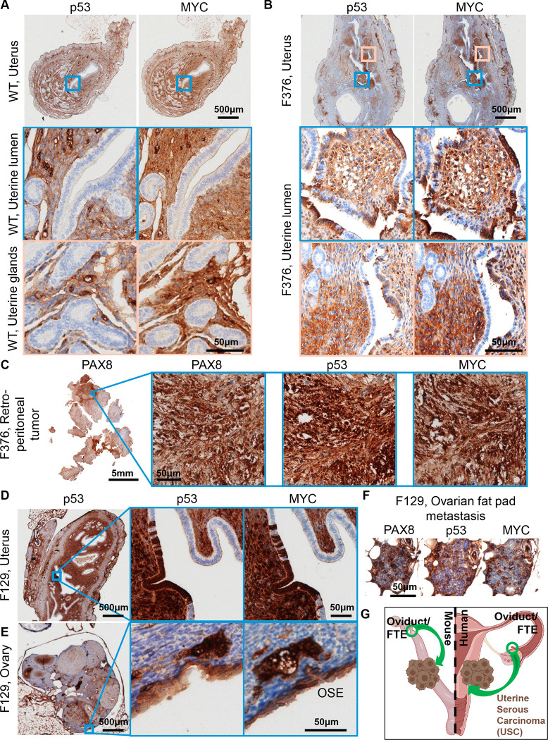
Figure 5. Uterine luminal epithelium staining of p53 and MYC.
(A) A wild-type mouse at 12mo of age shows negative p53 and MYC staining in uterine epithelium and positive staining in adjacent tissue. (B) Example of a 12mo old OvTrpMyc mouse with cytoplasmic p53 and whole-cell MYC staining along uterine luminal epithelium. (C) Large retroperitoneal tumor, behind kidney, stained for p53, MYC, and PAX8. (D) An example of uterine luminal epithelium expression in OvTrpMyc mouse F129, with evidence of similar whole-cell staining of p53 and MYC on the (E) OSE and (F) ovarian fat pad metastasis. Mouse F129 was 11mo of age. (G) Proposed model for observed dissemination of cells from FTE to uterine luminal epithelium as an origin of uterine serous carcinoma

