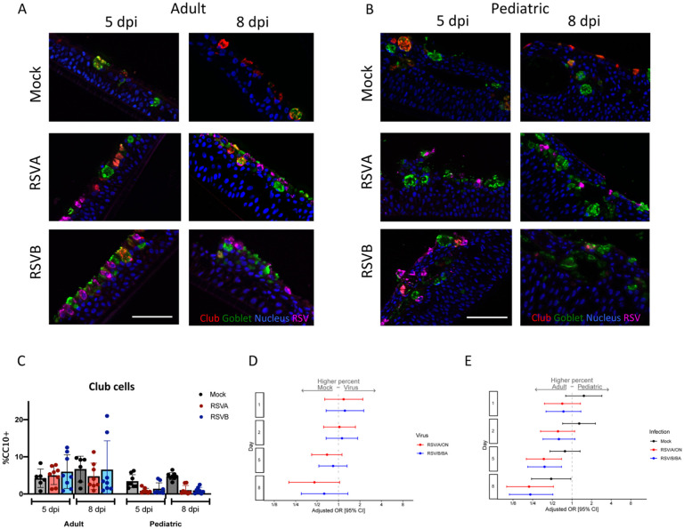
Figure 6. Club cells in pediatric and adult HNO-ALIs.
A) Representative IF images of a single adult HNO and B) single pediatric HNO at day 5 and 8 post infection. Club cells are stained red with CC10, goblet cells are stained in green by Muc5AC, RSV particles are stained in magenta by anti-RSV antibody, and cell nuclei are stained in blue by DAPI. Scale bar is 100 μm. C) Percentage of club cells (CC10+ cells with DAPI+ nuclei over total number of DAPI+ cells) in adult compared to pediatric HNO-ALIs at 5- and 8-day post-infection (dpi). D) Club cell percentage data was modeled using the following factors: the interaction of dpi and HNO age, the interaction of age and viral infection, and the interaction of dpi and virus. Adjusted odds ratio estimates with 95% confidence intervals are represented by dots and T-bars, respectively. E) Adjusted odds ratio with 95% confidence intervals of higher percentage of club cells in adult compared to pediatric HNOs.

