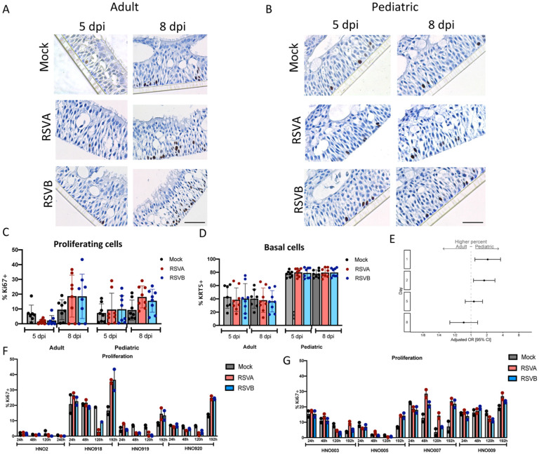
Figure 7: Cell proliferation in pediatric and adult HNO-ALIs.
A) Representative Ki67 staining of a single adult HNO-ALI and B) single pediatric HNO-ALI at 5- and 8-day post infection (dpi) with RSV/A/ON, RSV/B/BA, or mock infection. Scale bar is 100 μm. C) Percentage of proliferating cells (number of Ki67 positive cells/ total cells) in 4 adult HNO-ALIs versus 4 pediatric HNO-ALIs at 5 or 8 dpi after infection with RSV/A/ON, RSV/B/BA, or mock infection. D) Percentage of basal cells (number of Krt5 positive cells/ total cells) in 4 adult HNO-ALIs versus 4 pediatric HNO-ALIs at 5 or 8 dpi after infection with RSV/A/ON, RSV/B/BA, or mock infection. E) Ki67 percent data was modeled using the following factors: the interaction of dpi and HNO age as well as the interaction of dpi and virus. Forest plot showing the adjusted odds ratio with 95% confidence intervals between adult and pediatric HNOs of having a higher amount of proliferating cells. Adjusted odds ratio estimates and their associated 95% confidence intervals are represented by dots and T-bars, respectively. There was no difference between mock and RSV infected HNOs, and thus this was dropped from the model and graph. F) Percentage of proliferating cells, in 4 adult HNO-ALIs at 1, 2, 5, and 8 dpi with RSV/A/ON or RSV/B/BA or mock infection. Each line shows variability in amount of proliferation. G) Percentage of proliferating cells in 4 pediatric HNO-ALIs at 1, 2, 5, and 8 dpi with RSV/A/ON or RSV/B/BA or mock infection.

