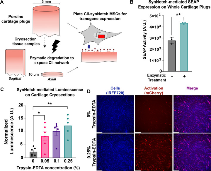
Figure 2: CII-synNotch MSCs detect degradation of cartilage tissue.
(A) 3 mm-diameter primary porcine cartilage explants were enzymatically treated or cryosectioned prior to enzymatic treatment. Enzyme treatment reveals CII epitopes detected by the mAbCII recognition motif. (B) SynNotch-inducible SEAP expression after plating engineered cells on enzymatically-damaged whole-plug explants (t=72 hr, Welch’s t-test on n=3 replicates per group). (C) Inducible luciferase expression from engineered cells on cryosections treated with varying concentrations of trypsin-EDTA (t=48hr). (D) Representative images of synNotch activation on sagittal cryosection samples without treatment and with 0.25% trypsin-EDTA (t=48hr). iRFP is false-colored as blue to enable reporter discrimination in the merged image. Scale = 200 µm. Data plotted as mean ± SEM. *p<0.05, **p<0.01.

