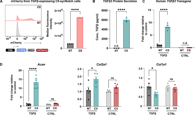
Figure 4: CII-synNotch effectively regulates pro-anabolic TGFβ3 expression.
(A) A synNotch payload contains human TGFβ3 co-expressed with mCherry, connected through an internal ribosomal entry site (IRES), allowing for expression of both transgenes. Inducible mCherry measurements of TGFβ3-expressing cells with and without activation on CII as a readout of synNotch activation (t=72 hours, Student’s t-test on n=3 replicates per group). (B) ELISA measurements of TGFβ3 after plating on CII-adsorption to cell culture surfaces (t=72 hours, Student’s t-test on n=3 replicates per group). (C) qRT-PCR detection of transgene hTGFβ3 in experiments comparing TGFβ3-expressing cells to reporter-expressing cells. qRT-PCR data is normalized to no-treatment controls for each cell line. (D) qRT-PCR detection of pro-anabolic Acan and Col2a1 expression, as well as Col1a1 expression. All qRT-PCR: t=72 hrs, two-way ANOVA with Tukey’s post hoc analysis on n=5 replicates per group. *p<0.05, ****p<0.0001. n.d.: not detected.

