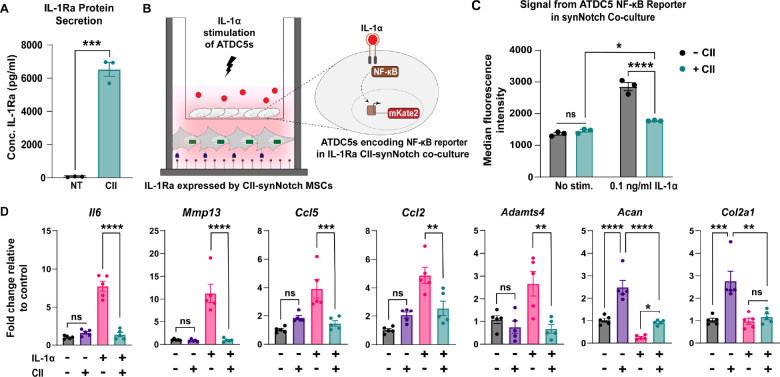
Figure 5: CII-synNotch MSCs mitigate IL-1-induced inflammation in a CII-dependent manner.
(A) ELISA measurements of IL-1Ra after plating on CII-decorated surfaces (t=72 hours, Student’s t-test on n=3 replicates per group). (B) Schematic of trans-well co-culture for assessment of IL-1Ra synNotch circuits on inflammatory chondrocytes. Bottom: 25 μg/ml of CII is plated for activation of CII-synNotch MSCs containing inducible IL-1Ra. Top: ATDC5 chondrocytes are plated in 3 μm-porous inserts. After three days of synNotch activation by MSCs, 0.1 ng/ml IL-1α was added to the insert. Flow cytometry and qRT-PCR results were obtained two days after stimulation. (C) ATDC5s were transduced with an mKate2 reporter that is upregulated in response to NF-κB-driven inflammation. Flow cytometry of mKate2 reports for the effect of inducible IL-1Ra expression on the NF-κB signal in IL-1-stimulated co-cultures (t=5d from plating, two-way ANOVA on n=3 replicates per group). (D) qRT-PCR detection of pro-inflammatory genes Il6, Mmp13, Ccl5, Ccl2, and Adamts4 as well as ECM-related Acan and Col2a1. All qRT-PCR data is normalized to chondrocyte gene expression in co-cultures with neither CII-synNotch activation nor IL-1α stimulation (t=5d from plating, two-way ANOVA on n=five replicates per group). *p<0.05, **p<0.01, ***p<0.001, ****p<0.0001.

