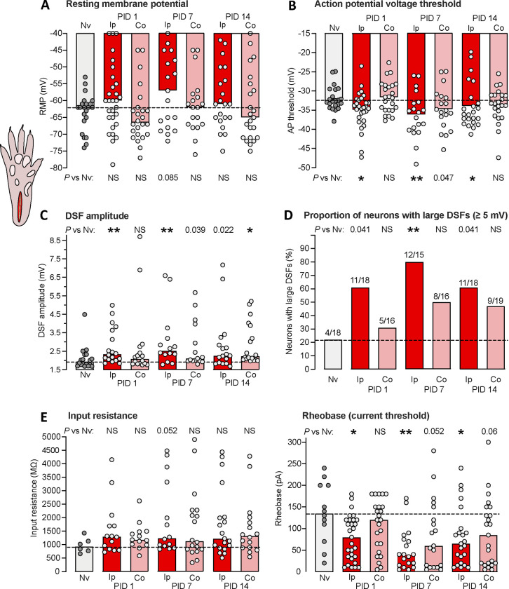
Figure 2.
Hyperexcitable alterations found in dissociated DRG neuron somata up to 2 weeks after unilateral plantar incision in vivo. (A) Lack of significant alterations in RMP. In all panels, bars represent medians or proportions; dashed line is the value for the naïve group. Points in a panel represent values for individual neurons. Comparisons by Mann-Whitney U tests with Bonferroni corrections were made for each time point versus naïve group performed separately for the ipsilateral and contralateral groups. Here and in all figures, any numeric P values <0.10 are reported to document possible trends. (B) Alterations in AP voltage threshold. Separate comparisons for ipsilateral and contralateral groups were made at each time point versus naïve group by Mann-Whitney U tests with Bonferroni corrections. Note that a numeric P value <0.05 reported in the figure means that the comparison did not meet statistical significance after Bonferroni correction. (C) Alterations in DSF amplitude, with separate comparisons made for ipsilateral and contralateral groups at each time point versus naïve group by Mann-Whitney U tests with Bonferroni corrections. (D) Alterations in the proportion of DRG neurons exhibiting large DSFs (≥ 5 mV). Separate comparisons were made for ipsilateral and contralateral groups at each time point versus naïve group by Fisher’s exact tests. (E) Lack of alterations in input resistance assessed by Mann-Whitney U tests with Bonferroni corrections. (F) Alterations in rheobase. Separate comparisons for ipsilateral and contralateral groups at each time point versus naïve group by Mann-Whitney U tests with Bonferroni corrections. AP, action potential; Co, contralateral to injury; DSF, depolarizing spontaneous fluctuation; Ip, ipsilateral; Nv, naïve; NS, not significant; PID, postinjury day.

