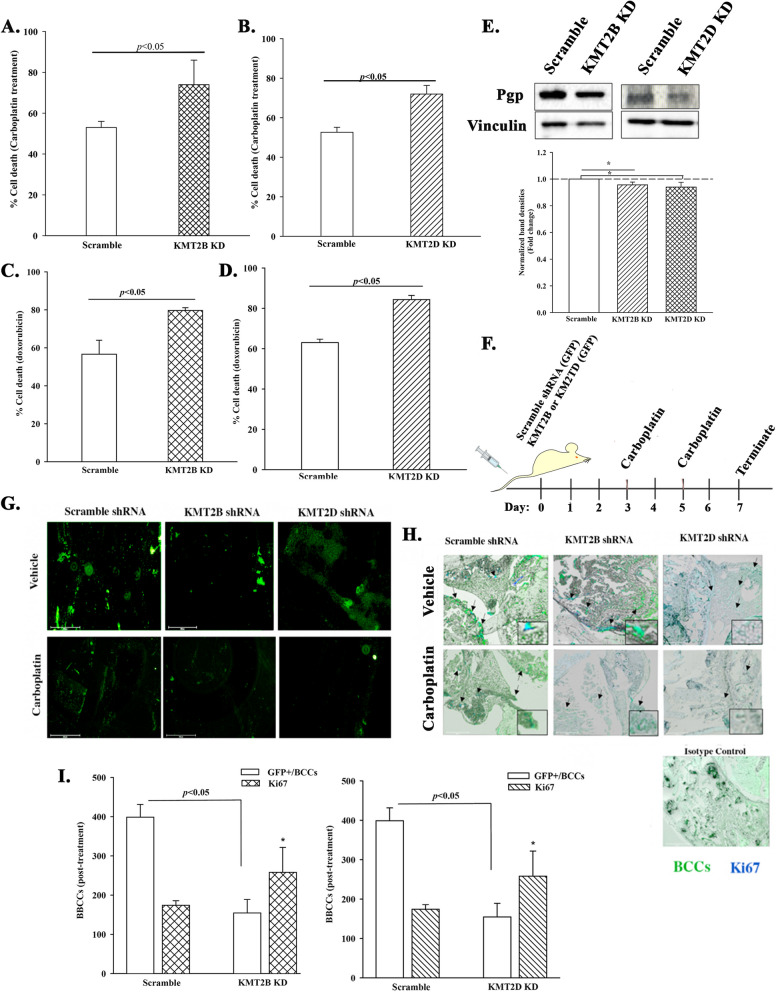
Fig. 6.
In vitro and in vivo chemosensitivity of KMT2B and KMT2D KD BCCs. A & B KMT2B (A) or KMT2D (B) KD BCCs were treated with 200 μg/mL carboplatin for 48 h. Control BCCs were transfected with scramble shRNA and similarly treated. The cells were analyzed with the MTT assay and the results presented as mean ± SD cell death for three biological replicates. Each replicate contained three technical studies. C & D The studies in `A’ were repeated except for treatment with doxorubicin (1 μM) for 48 h. E Western blot for P-gp with extracts from KMT2B and KMT2D KD BCCs, and BCCs with scramble shRNA. The lower graph showed the mean fold change±SD of normalized bands, n = 3. * p < .05. F Diagram showing the in vivo model in which KMT2B or KMT2D KD MDA-MB-231 BCCs were injected in the tail veins of 6-wk female nude mice. Control mice were injected with BCCs containing scramble shRNA. Shown are the timeline (days 3 and 5) treatment with carboplatin (5 mg/kg) or vehicle (1X PBS) on days 3 and 5. The studies were terminated at day 7. G Femurs from the euthanized mice in `F′ were scaped at the endosteal region and then immediately examined on an Evos Fl2 for GPF-positive BCCs. Shown are representative images for five femurs, each from a different mouse. H Femurs from the mice described in F and G were decalcified and embedded in paraffin. Sections were analyzed on the Evos Fl2 for GFP (green) or labeled for Ki67 (blue) (teal cells = green (BCCs) + blue (Ki67). Shown are representative image at 10X magnification. I Figures show the total number of GFP+ cells in 10 fields of sections from mouse femurs and KI67+ cells within the GFP+ cells. * p < .05 vs. scramble Ki67

