Fig. 4.
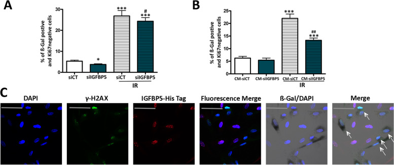
Paracrine action of IGFBP5. A MSCs transfected with control siRNA or IGFBP5-siRNA were X-ray irradiated, and senescence was evaluated 48 h later. The graph depicts the percentage of senescent cells under different experimental conditions. The symbols *** p < 0.001 and * p < 0.05 indicate statistical significance between the control (siCT) and other samples. The # (p < 0.05) indicateds statistical significance between irradiated siCT versus irradiated siIGFBP5 (B) Healthy MSCs were incubated for 48 h with conditioned media (CM) obtained from the previously mentioned samples. The graph illustrates the percentage of senescent cells under different experimental conditions. The symbol *** p < 0.001 indicates statistical significance between the control (siCT) and other samples. The ## (p < 0.01) indicateds statistical significance between irradiated siCT versus irradiated siIGFBP5. C Representative images of cells stained with anti-γH2AX (green) and His-tag IGFBP5 (red) are shown. Cell nuclei were stained with DAPI. The β-galactosidase activity was evidenced as dark gray. We employed a Leica CTR500 microscope, which was equipped with a DCF3000G digital monochrome camera. The β-galactosidase activity was captured as a gray-stain using this configuration. The arrows show cells that were β-galactosidase/γH2AX positive and IGFBP5 negative

