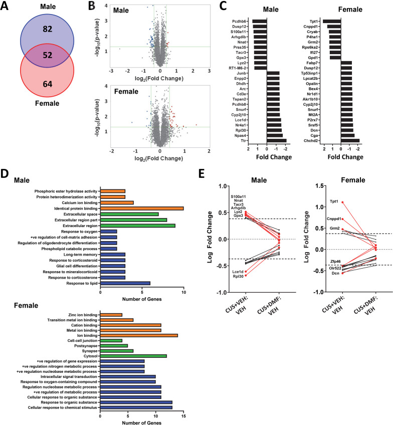
Fig. 4.
Sex-specific alterations in hippocampal gene expression in response to CUS and the mitigating effects of DMF. A The Venn diagram depicts the number of transcripts with a log2 fold change ≥ 0.4 or ≤ − 0.4 in males (blue) and females (red) following CUS. B Volcano plots represent the -log10(p-value) against the log2(fold change) magnitude of transcripts altered by CUS in males (top panel) and females (bottom panel). C The top 24 differentially expressed genes for each sex following CUS. D Gene Ontology analysis identified significantly enriched terms from males (top panel) and females (bottom panel) in the categories of molecular function (orange), cellular components (green), and biological processes (blue). E DMF normalization of CUS-induced changes in gene expression, focusing on transcripts with a minimal log2 fold correction of ≥ 0.3 or ≤ − 0.3

